Event submissions
Published
 10.3390/CPPC2021-10221 (registering DOI)
 This submission belongs to the session A. Chemical-Biology and Medicinal Chemistry of the event First Canadian Peptide and Protein Community Virtual Symposium
Published date
27 May, 2021
Citation
Melanie Forêt Jacquard, Benoit Gautier, Baptiste Legrand, Nicolas Tricaud, Nicolas Inguimbert, Mitochondrial VDAC1 Pporin as a Therapeutic Target in Demyelination Process: Investigation of the Interaction Sites between Hexokinase I and VDAC1, in Proceedings of First Canadian Peptide and Protein Community Virtual Symposium, 27 May–28 May 2021, MDPI: Basel, Switzerland, doi: 10.3390/CPPC2021-10221
Share
Email
Facebook
Twitter
LinkedIn
Mitochondrial VDAC1 Pporin as a Therapeutic Target in Demyelination Process: Investigation of the Interaction Sites between Hexokinase I and VDAC1
'%3e%3cpath%20d='M12.6647%2010.9104C12.4176%2010.325%2012.0589%209.7933%2011.6088%209.34485C11.16%208.89511%2010.6283%208.53653%2010.0432%208.28891C10.038%208.28629%2010.0327%208.28498%2010.0275%208.28236C10.8437%207.69282%2011.3743%206.73252%2011.3743%205.64907C11.3743%203.85423%209.92005%202.40002%208.12522%202.40002C6.33038%202.40002%204.87618%203.85423%204.87618%205.64907C4.87618%206.73252%205.40677%207.69282%206.22296%208.28367C6.21772%208.28629%206.21248%208.2876%206.20724%208.29022C5.62031%208.53783%205.09365%208.89287%204.64167%209.34616C4.19192%209.79496%203.83334%2010.3266%203.58573%2010.9117C3.34247%2011.4846%203.21128%2012.0987%203.19925%2012.721C3.1989%2012.735%203.20135%2012.7489%203.20646%2012.7619C3.21157%2012.7749%203.21924%2012.7868%203.22901%2012.7968C3.23877%2012.8068%203.25045%2012.8148%203.26334%2012.8202C3.27623%2012.8256%203.29007%2012.8284%203.30406%2012.8284H4.09012C4.14776%2012.8284%204.19362%2012.7825%204.19493%2012.7262C4.22113%2011.7148%204.62726%2010.7676%205.34519%2010.0497C6.08802%209.30686%207.07452%208.89811%208.12522%208.89811C9.17592%208.89811%2010.1624%209.30686%2010.9052%2010.0497C11.6232%2010.7676%2012.0293%2011.7148%2012.0555%2012.7262C12.0568%2012.7839%2012.1027%2012.8284%2012.1603%2012.8284H12.9464C12.9604%2012.8284%2012.9742%2012.8256%2012.9871%2012.8202C13%2012.8148%2013.0117%2012.8068%2013.0214%2012.7968C13.0312%2012.7868%2013.0389%2012.7749%2013.044%2012.7619C13.0491%2012.7489%2013.0515%2012.735%2013.0512%2012.721C13.0381%2012.0947%2012.9084%2011.4856%2012.6647%2010.9104ZM8.12522%207.90243C7.52388%207.90243%206.95792%207.66793%206.53214%207.24215C6.10636%206.81636%205.87185%206.2504%205.87185%205.64907C5.87185%205.04773%206.10636%204.48177%206.53214%204.05599C6.95792%203.63021%207.52388%203.3957%208.12522%203.3957C8.72655%203.3957%209.29252%203.63021%209.7183%204.05599C10.1441%204.48177%2010.3786%205.04773%2010.3786%205.64907C10.3786%206.2504%2010.1441%206.81636%209.7183%207.24215C9.29252%207.66793%208.72655%207.90243%208.12522%207.90243Z'%20fill='%235D1EE1'/%3e%3c/g%3e%3c/svg%3e)
Melanie Forêt Jacquard 1
Benoit Gautier 2
Baptiste Legrand 3
Nicolas Tricaud 2
Nicolas Inguimbert 1
1. [a] Centre de Recherche Insulaire et Observatoire de l’Environnement (CRIOBE), USR CNRS 3278, CNRS-EPHE-UPVD, Université de Perpignan Via Domitia, Bâtiment T, 58 Avenue P. Alduy, 66860 Perpignan, France.
2. [b] Institut des Neurosciences de Montpellier, INSERM U1051, Université de Montpellier, 80 Rue A. Fliche, 34091 Montpellier, France.
3. [c] Institut des Biomolécules Max Mousseron, UMR 5247, CNRS, UM, ENSCM, UFR des Sciences Pharmaceutiques et Biologiques, 15 Avenue Charles Flahault, 34093 Montpellier Cedex 5, France.
Abstract
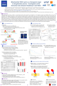
Mitochondrial dysfunction has been implicated in many diseases including cancer, cardiovascular and neurodegenerative diseases1,2. The voltage-dependant anion channel 1 (VDAC1) is the most abundant protein found in the outer mitochondrial membrane. VDAC1 functions as a gatekeeper and is considered as a multi-functional porin involved in cell survival and cell death3. VDAC1 mediates energy production as well as ions and metabolic exchanges between the mitochondria and other cellular compartments, thus modulating mitochondrial permeability. VDAC1 is also involved in the cellular Ca2+ homeostasis by its capacity to transport Ca2+ in and out of mitochondria4. In addition, VDAC1 interacts with multiple proteins implicated in neurodegeneration process5 as well as with pro-apoptotic and anti-apoptotic regulating proteins, and in particular Hexokinase isoforms I and II, its main ligand.
Hexokinase I (HK I) is found in brain tissues and is known as the “guardian of the mitochondria”. HK possess a hydrophobic N-terminal structured in α-helix that is necessary for the binding of HK to VDAC6. The interaction between HK and VDAC1 has attracted much interest, and several studies have proposed different models of binding to VDAC17,8. However, the precise mechanism of binding remains undefined up to now.
In order to give some insights to the nature of HK binding to VDAC1, we have developed new peptide analogs of HK I based on Hexokinase I N-terminal sequence. In this study, the in-vitro activity of the peptides was studied, the peptide helical content was investigated by circular dichroism and NMR, and peptide proteolytic stability was assessed.
(1) Shoshan-Barmatz, V.; Shteinfer-Kuzmine, A.; Verma, A. VDAC1 at the Intersection of Cell Metabolism, Apoptosis, and Diseases. Biomolecules 2020, 10 (11). https://doi.org/10.3390/biom10111485.
(2) Abramov, A. Y.; Berezhnov, A. V.; Fedotova, E. I.; Zinchenko, V. P.; Dolgacheva, L. P. Interaction of Misfolded Proteins and Mitochondria in Neurodegenerative Disorders. Biochem Soc Trans 2017, 45 (4), 1025–1033. https://doi.org/10.1042/BST20170024.
(3) Shoshan-Barmatz, V.; Ben-Hail, D. VDAC, a Multi-Functional Mitochondrial Protein as a Pharmacological Target. Mitochondrion 2012, 12 (1), 24–34. https://doi.org/10.1016/j.mito.2011.04.001.
(4) Shoshan-Barmatz, V.; Krelin, Y.; Shteinfer-Kuzmine, A. VDAC1 Functions in Ca2+ Homeostasis and Cell Life and Death in Health and Disease. Cell Calcium 2018, 69, 81–100. https://doi.org/10.1016/j.ceca.2017.06.007.
(5) Magri, A.; Messina, A. Interactions of VDAC with Proteins Involved in Neurodegenerative Aggregation: An Opportunity for Advancement on Therapeutic Molecules. Curr Med Chem 2017, 24 (40), 4470–4487. https://doi.org/10.2174/0929867324666170601073920.
(6) Xie, G. C.; Wilson, J. E. Rat Brain Hexokinase: The Hydrophobic N-Terminus of the Mitochondrially Bound Enzyme Is Inserted in the Lipid Bilayer. Arch Biochem Biophys 1988, 267 (2), 803–810. https://doi.org/10.1016/0003-9861(88)90090-2.
(7) Rosano, C. Molecular Model of Hexokinase Binding to the Outer Mitochondrial Membrane Porin (VDAC1): Implication for the Design of New Cancer Therapies. Mitochondrion 2011, 11 (3), 513–519. https://doi.org/10.1016/j.mito.2011.01.012.
(8) Abu-Hamad, S.; Zaid, H.; Israelson, A.; Nahon, E.; Shoshan-Barmatz, V. Hexokinase-I Protection against Apoptotic Cell Death Is Mediated via Interaction with the Voltage-Dependent Anion Channel-1: MAPPING THE SITE OF BINDING. J. Biol. Chem. 2008, 283 (19), 13482–13490. https://doi.org/10.1074/jbc.M708216200.
Keywords
mitochondria
VDAC1
Hexokinase
neurodegeneration
demyelination
Ca2+ homeostasis
peptides
Manuscript
sciforum-045399.pdf
Poster
2021-06-10 Poster CPPC 2021_modif_FORET JACQUARD Melanie 045399.pdf
Identification of Allergenic Epitopes in the Sequences of Rapeseed Seed Proteins
Effects Of The Cowpea Gln-Asp-Phe Peptide Daily Administration in Rats Fed A Saturated High-Fat Diet