Event submissions
Published
 10.3390/CPPC2021-10239 (registering DOI)
 This submission belongs to the session C. Structural Biology and Computational Methods of the event First Canadian Peptide and Protein Community Virtual Symposium
Published date
27 May, 2021
Citation
Ivonne Rebeca Lopez-Miranda, Dennis D. Fernandes, Omar Almurad, Yevgen Moskalenko, R. Scott Prosser, Claudiu C. Gradinaru, Measuring Conformational Selection in G Protein-Coupled Receptors by Single-Molecule Förster Resonance Energy Transfer, in Proceedings of First Canadian Peptide and Protein Community Virtual Symposium, 27 May–28 May 2021, MDPI: Basel, Switzerland, doi: 10.3390/CPPC2021-10239
Share
Email
Facebook
Twitter
LinkedIn
Measuring Conformational Selection in G Protein-Coupled Receptors by Single-Molecule Förster Resonance Energy Transfer
'%3e%3cpath%20d='M12.6647%2010.9104C12.4176%2010.325%2012.0589%209.7933%2011.6088%209.34485C11.16%208.89511%2010.6283%208.53653%2010.0432%208.28891C10.038%208.28629%2010.0327%208.28498%2010.0275%208.28236C10.8437%207.69282%2011.3743%206.73252%2011.3743%205.64907C11.3743%203.85423%209.92005%202.40002%208.12522%202.40002C6.33038%202.40002%204.87618%203.85423%204.87618%205.64907C4.87618%206.73252%205.40677%207.69282%206.22296%208.28367C6.21772%208.28629%206.21248%208.2876%206.20724%208.29022C5.62031%208.53783%205.09365%208.89287%204.64167%209.34616C4.19192%209.79496%203.83334%2010.3266%203.58573%2010.9117C3.34247%2011.4846%203.21128%2012.0987%203.19925%2012.721C3.1989%2012.735%203.20135%2012.7489%203.20646%2012.7619C3.21157%2012.7749%203.21924%2012.7868%203.22901%2012.7968C3.23877%2012.8068%203.25045%2012.8148%203.26334%2012.8202C3.27623%2012.8256%203.29007%2012.8284%203.30406%2012.8284H4.09012C4.14776%2012.8284%204.19362%2012.7825%204.19493%2012.7262C4.22113%2011.7148%204.62726%2010.7676%205.34519%2010.0497C6.08802%209.30686%207.07452%208.89811%208.12522%208.89811C9.17592%208.89811%2010.1624%209.30686%2010.9052%2010.0497C11.6232%2010.7676%2012.0293%2011.7148%2012.0555%2012.7262C12.0568%2012.7839%2012.1027%2012.8284%2012.1603%2012.8284H12.9464C12.9604%2012.8284%2012.9742%2012.8256%2012.9871%2012.8202C13%2012.8148%2013.0117%2012.8068%2013.0214%2012.7968C13.0312%2012.7868%2013.0389%2012.7749%2013.044%2012.7619C13.0491%2012.7489%2013.0515%2012.735%2013.0512%2012.721C13.0381%2012.0947%2012.9084%2011.4856%2012.6647%2010.9104ZM8.12522%207.90243C7.52388%207.90243%206.95792%207.66793%206.53214%207.24215C6.10636%206.81636%205.87185%206.2504%205.87185%205.64907C5.87185%205.04773%206.10636%204.48177%206.53214%204.05599C6.95792%203.63021%207.52388%203.3957%208.12522%203.3957C8.72655%203.3957%209.29252%203.63021%209.7183%204.05599C10.1441%204.48177%2010.3786%205.04773%2010.3786%205.64907C10.3786%206.2504%2010.1441%206.81636%209.7183%207.24215C9.29252%207.66793%208.72655%207.90243%208.12522%207.90243Z'%20fill='%235D1EE1'/%3e%3c/g%3e%3c/svg%3e)
Ivonne Rebeca Lopez-Miranda 1,2
Dennis D. Fernandes 1,3
Omar Almurad 1
Yevgen Moskalenko 1,3
R. Scott Prosser 4,5
Claudiu C. Gradinaru 1,6
1. Department of Chemical & Physical Sciences, University of Toronto Mississauga, Mississauga, Ontario, L5L 1C6, Canada
2. Department of Economics, University of Toronto Mississauga, Mississauga, Ontario, L5L 1C6, Canada
3. Department of Physics, University of Toronto, Ontario, M5S 1A7, Canada
4. Department of Chemical & Physical Sciences, University of Toronto Mississauga, Mississauga, Ontario, L5L 1C6, Canada.
5. Department of Biochemistry, University of Toronto, 1 King’s College Circle, Toronto, Ontario M5S 1A8, Canada
6. Department of Physics, University of Toronto, Toronto, Ontario, M5S 1A7, Canada
Abstract
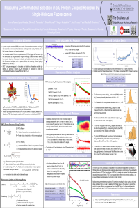
Fluorescent probes and techniques can be used to study protein structures. Molecular processes that occur in living organisms are visualized through the use of fluorescence techniques, which are sensitive enough to detect miniscule changes. The intracellular details of G protein-coupled receptors (GPCRs) tagged with fluorescent dyes can be visualized by using fluorescence imaging microscopy. Confocal microscopy can detect sub-cellular structures, and by combining Forster resonance energy transfer (FRET) techniques with fluorescence microscopy, one can determine physical molecular interactions within proteins. By using fluorescence spectroscopy, spatial information regarding the molecular behaviour of fluorescent molecules can be determined by using a variety of fluorescence parameters: quantum yield, which relates to intensity, spectrum, lifetime, and anisotropy. These techniques are extremely sensitive to changes and minimally invasive. FRET techniques were applied to characterize and detail the conformational changes exhibited by the A2A Adenosine receptor reconstituted in HDL nanodiscs, labelled with Alexa 488 and Alexa 647 dyes at the T119C and Q226C positions on transmembrane helices 4 and 6. A variety of ligand conditions would induce unique separation distances between these helices, resulting in FRET efficiency values which correspond to distinct conformations of the receptor. Specifically, single-molecule Forster resonance energy transfer (smFRET) was performed on the receptor to distinguish active and inactive states, pertaining to different FRET efficiency values. Characterization of the sample involved using many fluorescence techniques, such as fluorescence correlation spectroscopy (FCS), emission and excitation spectra, fluorescence anisotropy decay (FAD), and fluorescence lifetime.
Keywords
GPCRs
single-molecule fluorescence
smFRET
FCS
mini-G
A2A Adenosine Receptor
Manuscript
sciforum-045252.pdf
Poster
sciforum_045252_slides_IvonneLopezMiranda_Poster_smFRET_A2A_Adenosine_Receptor_1.pdf
Targeting SARS-COV-2 Receptor Binding Domain with Stapled Peptides: An In-Silico Study
Supramolecular Ultra-Short Dehydropeptide-Based Hydrogels As Potential Affordable Nanocarriers