Event submissions
Published

This submission belongs to the session 5. Developmental Neuroscience of the event The 2nd International Electronic Conference on Brain Sciences
Published date
14 Jul, 2021
Academic Editor
'%3e%3cpath%20d='M12.6657%2010.9104C12.4185%2010.325%2012.0599%209.79327%2011.6097%209.34482C11.1609%208.89508%2010.6293%208.5365%2010.0442%208.28888C10.0389%208.28626%2010.0337%208.28495%2010.0285%208.28233C10.8446%207.69279%2011.3752%206.73249%2011.3752%205.64904C11.3752%203.8542%209.92103%202.39999%208.1262%202.39999C6.33136%202.39999%204.87715%203.8542%204.87715%205.64904C4.87715%206.73249%205.40774%207.69279%206.22393%208.28364C6.21869%208.28626%206.21345%208.28757%206.20821%208.29019C5.62129%208.5378%205.09463%208.89284%204.64265%209.34613C4.1929%209.79493%203.83432%2010.3266%203.58671%2010.9117C3.34345%2011.4845%203.21226%2012.0987%203.20023%2012.7209C3.19988%2012.7349%203.20233%2012.7488%203.20744%2012.7619C3.21255%2012.7749%203.22022%2012.7867%203.22998%2012.7968C3.23975%2012.8068%203.25142%2012.8147%203.26431%2012.8202C3.2772%2012.8256%203.29105%2012.8284%203.30504%2012.8284H4.09109C4.14874%2012.8284%204.19459%2012.7825%204.1959%2012.7262C4.2221%2011.7148%204.62823%2010.7676%205.34617%2010.0497C6.08899%209.30683%207.0755%208.89808%208.1262%208.89808C9.17689%208.89808%2010.1634%209.30683%2010.9062%2010.0497C11.6242%2010.7676%2012.0303%2011.7148%2012.0565%2012.7262C12.0578%2012.7838%2012.1037%2012.8284%2012.1613%2012.8284H12.9474C12.9613%2012.8284%2012.9752%2012.8256%2012.9881%2012.8202C13.001%2012.8147%2013.0126%2012.8068%2013.0224%2012.7968C13.0322%2012.7867%2013.0398%2012.7749%2013.0449%2012.7619C13.0501%2012.7488%2013.0525%2012.7349%2013.0522%2012.7209C13.0391%2012.0947%2012.9094%2011.4855%2012.6657%2010.9104ZM8.1262%207.9024C7.52486%207.9024%206.9589%207.6679%206.53312%207.24211C6.10733%206.81633%205.87283%206.25037%205.87283%205.64904C5.87283%205.0477%206.10733%204.48174%206.53312%204.05596C6.9589%203.63018%207.52486%203.39567%208.1262%203.39567C8.72753%203.39567%209.29349%203.63018%209.71927%204.05596C10.1451%204.48174%2010.3796%205.0477%2010.3796%205.64904C10.3796%206.25037%2010.1451%206.81633%209.71927%207.24211C9.29349%207.6679%208.72753%207.9024%208.1262%207.9024Z'%20fill='%235D1EE1'/%3e%3c/g%3e%3c/svg%3e) Stephen Meriney
Stephen MerineyCitation
Polina Shkorbatova, Vsevolod Lyakhovetskii, Svetlana Alexeenko, Density of SMI-32 immunopositive neurons in eye-specific layers of lateral geniculate nucleus in kittens reared with monocular deprivation and unilateral convergent squint., in Proceedings of The 2nd International Electronic Conference on Brain Sciences, 15 July–30 July 2021, MDPI: Basel, Switzerland, doi: 10.3390/IECBS2021-10666
Share
Email
Facebook
Twitter
LinkedIn
Density of SMI-32 immunopositive neurons in eye-specific layers of lateral geniculate nucleus in kittens reared with monocular deprivation and unilateral convergent squint.

Polina Shkorbatova 1

Vsevolod Lyakhovetskii 2
'%3e%3cpath%20d='M12.6647%2010.9104C12.4176%2010.325%2012.0589%209.7933%2011.6088%209.34485C11.16%208.89511%2010.6283%208.53653%2010.0432%208.28891C10.038%208.28629%2010.0327%208.28498%2010.0275%208.28236C10.8437%207.69282%2011.3743%206.73252%2011.3743%205.64907C11.3743%203.85423%209.92005%202.40002%208.12522%202.40002C6.33038%202.40002%204.87618%203.85423%204.87618%205.64907C4.87618%206.73252%205.40677%207.69282%206.22296%208.28367C6.21772%208.28629%206.21248%208.2876%206.20724%208.29022C5.62031%208.53783%205.09365%208.89287%204.64167%209.34616C4.19192%209.79496%203.83334%2010.3266%203.58573%2010.9117C3.34247%2011.4846%203.21128%2012.0987%203.19925%2012.721C3.1989%2012.735%203.20135%2012.7489%203.20646%2012.7619C3.21157%2012.7749%203.21924%2012.7868%203.22901%2012.7968C3.23877%2012.8068%203.25045%2012.8148%203.26334%2012.8202C3.27623%2012.8256%203.29007%2012.8284%203.30406%2012.8284H4.09012C4.14776%2012.8284%204.19362%2012.7825%204.19493%2012.7262C4.22113%2011.7148%204.62726%2010.7676%205.34519%2010.0497C6.08802%209.30686%207.07452%208.89811%208.12522%208.89811C9.17592%208.89811%2010.1624%209.30686%2010.9052%2010.0497C11.6232%2010.7676%2012.0293%2011.7148%2012.0555%2012.7262C12.0568%2012.7839%2012.1027%2012.8284%2012.1603%2012.8284H12.9464C12.9604%2012.8284%2012.9742%2012.8256%2012.9871%2012.8202C13%2012.8148%2013.0117%2012.8068%2013.0214%2012.7968C13.0312%2012.7868%2013.0389%2012.7749%2013.044%2012.7619C13.0491%2012.7489%2013.0515%2012.735%2013.0512%2012.721C13.0381%2012.0947%2012.9084%2011.4856%2012.6647%2010.9104ZM8.12522%207.90243C7.52388%207.90243%206.95792%207.66793%206.53214%207.24215C6.10636%206.81636%205.87185%206.2504%205.87185%205.64907C5.87185%205.04773%206.10636%204.48177%206.53214%204.05599C6.95792%203.63021%207.52388%203.3957%208.12522%203.3957C8.72655%203.3957%209.29252%203.63021%209.7183%204.05599C10.1441%204.48177%2010.3786%205.04773%2010.3786%205.64907C10.3786%206.2504%2010.1441%206.81636%209.7183%207.24215C9.29252%207.66793%208.72655%207.90243%208.12522%207.90243Z'%20fill='%235D1EE1'/%3e%3c/g%3e%3c/svg%3e)
Svetlana Alexeenko 3
1. Neuromorphology laboratory, Pavlov Institute of Physiology RAS
2. Motor physiology laboratory, Pavlov Institute of Physiology RAS
3. Vision physiology, Pavlov Institute of Physiology RAS
Abstract
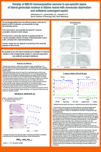
The non-phosphorylated heavy neurofilament proteins, which can be labelled by SMI-32 antibodies, are characteristic for large, fast-conducting neurons. In the visual system, such properties are typical for Y-neurons. It is known that in monocularly deprived or squinting animals the Y-neurons population declines in A-layers of lateral geniculate nucleus (LGNd), connected with a deprived/squinting eye. However if this loss depends on eccentricity of the visual field projection is still not known. To study this, as well as the time course of the developmental changes, we used the frontal sections of LGNd of both hemispheres of 2- and 3- month kittens reared with monocular deprivation or unilateral convergent squint. We had developed the custom software to divide the binocular part of A-layers into 10 consecutive sectors and to calculate the number of SMI-32 immunopositive neurons in each of them. The neuronal density was calculated and compared between groups in sectors with the same eccentricity. In monocularly deprived animals, decline of the neuronal density relative to the control group was found in layers, innervated from the deprived eye in both age groups, regardless of eccentricity. However in strabismic kittens the decrease in neuronal density was revealed only in the peripheral sectors of layer A1, driven by deviated eye. The width of this area of reduced Y-neuron density was larger in 3 months kittens, indicating that the development of the disorder has not yet stabilized. The results may be interpreted as morpho-physiological correlates of different types of human amblyopia.
Keywords
amblyopia
monocular deprivation
squint
development
Manuscript
sciforum-045642-done.pdf
Poster
poster LGN SMI32-4.pdf
Systematic review: Antioxidant and neuroprotective capacity of species of the genus Asplenium (Monilophyta: Aspleniaceae)
Comparing brain responses to different styles of music through their real and imagined interpretation: an analysis based on EEG connectivity networks