Event submissions
Published
 10.3390/ecsa-8-11305 (registering DOI)
 This submission belongs to the session P. Posters of the event 8th International Electronic Conference on Sensors and Applications
Published date
01 Nov, 2021
Academic Editor
'%3e%3cpath%20d='M12.6657%2010.9104C12.4185%2010.325%2012.0599%209.79327%2011.6097%209.34482C11.1609%208.89508%2010.6293%208.5365%2010.0442%208.28888C10.0389%208.28626%2010.0337%208.28495%2010.0285%208.28233C10.8446%207.69279%2011.3752%206.73249%2011.3752%205.64904C11.3752%203.8542%209.92103%202.39999%208.1262%202.39999C6.33136%202.39999%204.87715%203.8542%204.87715%205.64904C4.87715%206.73249%205.40774%207.69279%206.22393%208.28364C6.21869%208.28626%206.21345%208.28757%206.20821%208.29019C5.62129%208.5378%205.09463%208.89284%204.64265%209.34613C4.1929%209.79493%203.83432%2010.3266%203.58671%2010.9117C3.34345%2011.4845%203.21226%2012.0987%203.20023%2012.7209C3.19988%2012.7349%203.20233%2012.7488%203.20744%2012.7619C3.21255%2012.7749%203.22022%2012.7867%203.22998%2012.7968C3.23975%2012.8068%203.25142%2012.8147%203.26431%2012.8202C3.2772%2012.8256%203.29105%2012.8284%203.30504%2012.8284H4.09109C4.14874%2012.8284%204.19459%2012.7825%204.1959%2012.7262C4.2221%2011.7148%204.62823%2010.7676%205.34617%2010.0497C6.08899%209.30683%207.0755%208.89808%208.1262%208.89808C9.17689%208.89808%2010.1634%209.30683%2010.9062%2010.0497C11.6242%2010.7676%2012.0303%2011.7148%2012.0565%2012.7262C12.0578%2012.7838%2012.1037%2012.8284%2012.1613%2012.8284H12.9474C12.9613%2012.8284%2012.9752%2012.8256%2012.9881%2012.8202C13.001%2012.8147%2013.0126%2012.8068%2013.0224%2012.7968C13.0322%2012.7867%2013.0398%2012.7749%2013.0449%2012.7619C13.0501%2012.7488%2013.0525%2012.7349%2013.0522%2012.7209C13.0391%2012.0947%2012.9094%2011.4855%2012.6657%2010.9104ZM8.1262%207.9024C7.52486%207.9024%206.9589%207.6679%206.53312%207.24211C6.10733%206.81633%205.87283%206.25037%205.87283%205.64904C5.87283%205.0477%206.10733%204.48174%206.53312%204.05596C6.9589%203.63018%207.52486%203.39567%208.1262%203.39567C8.72753%203.39567%209.29349%203.63018%209.71927%204.05596C10.1451%204.48174%2010.3796%205.0477%2010.3796%205.64904C10.3796%206.25037%2010.1451%206.81633%209.71927%207.24211C9.29349%207.6679%208.72753%207.9024%208.1262%207.9024Z'%20fill='%235D1EE1'/%3e%3c/g%3e%3c/svg%3e) Stefano Mariani
Stefano MarianiCitation
Roland Hager, Peter Lanzerstorfer, Julian Weghuber, Christian Forsich, Applicability of polymeric substrates for subcellular live cell micropatterning experiments, in Proceedings of 8th International Electronic Conference on Sensors and Applications, 1 November–15 November 2021, MDPI: Basel, Switzerland, doi: 10.3390/ecsa-8-11305
Share
Email
Facebook
Twitter
LinkedIn
Applicability of polymeric substrates for subcellular live cell micropatterning experiments
'%3e%3cpath%20d='M12.6647%2010.9104C12.4176%2010.325%2012.0589%209.7933%2011.6088%209.34485C11.16%208.89511%2010.6283%208.53653%2010.0432%208.28891C10.038%208.28629%2010.0327%208.28498%2010.0275%208.28236C10.8437%207.69282%2011.3743%206.73252%2011.3743%205.64907C11.3743%203.85423%209.92005%202.40002%208.12522%202.40002C6.33038%202.40002%204.87618%203.85423%204.87618%205.64907C4.87618%206.73252%205.40677%207.69282%206.22296%208.28367C6.21772%208.28629%206.21248%208.2876%206.20724%208.29022C5.62031%208.53783%205.09365%208.89287%204.64167%209.34616C4.19192%209.79496%203.83334%2010.3266%203.58573%2010.9117C3.34247%2011.4846%203.21128%2012.0987%203.19925%2012.721C3.1989%2012.735%203.20135%2012.7489%203.20646%2012.7619C3.21157%2012.7749%203.21924%2012.7868%203.22901%2012.7968C3.23877%2012.8068%203.25045%2012.8148%203.26334%2012.8202C3.27623%2012.8256%203.29007%2012.8284%203.30406%2012.8284H4.09012C4.14776%2012.8284%204.19362%2012.7825%204.19493%2012.7262C4.22113%2011.7148%204.62726%2010.7676%205.34519%2010.0497C6.08802%209.30686%207.07452%208.89811%208.12522%208.89811C9.17592%208.89811%2010.1624%209.30686%2010.9052%2010.0497C11.6232%2010.7676%2012.0293%2011.7148%2012.0555%2012.7262C12.0568%2012.7839%2012.1027%2012.8284%2012.1603%2012.8284H12.9464C12.9604%2012.8284%2012.9742%2012.8256%2012.9871%2012.8202C13%2012.8148%2013.0117%2012.8068%2013.0214%2012.7968C13.0312%2012.7868%2013.0389%2012.7749%2013.044%2012.7619C13.0491%2012.7489%2013.0515%2012.735%2013.0512%2012.721C13.0381%2012.0947%2012.9084%2011.4856%2012.6647%2010.9104ZM8.12522%207.90243C7.52388%207.90243%206.95792%207.66793%206.53214%207.24215C6.10636%206.81636%205.87185%206.2504%205.87185%205.64907C5.87185%205.04773%206.10636%204.48177%206.53214%204.05599C6.95792%203.63021%207.52388%203.3957%208.12522%203.3957C8.72655%203.3957%209.29252%203.63021%209.7183%204.05599C10.1441%204.48177%2010.3786%205.04773%2010.3786%205.64907C10.3786%206.2504%2010.1441%206.81636%209.7183%207.24215C9.29252%207.66793%208.72655%207.90243%208.12522%207.90243Z'%20fill='%235D1EE1'/%3e%3c/g%3e%3c/svg%3e)
Roland Hager 1
Peter Lanzerstorfer 1
Julian Weghuber 1
Christian Forsich 1
1. University of Applied Sciences Upper Austria, Wels, Austria
Abstract
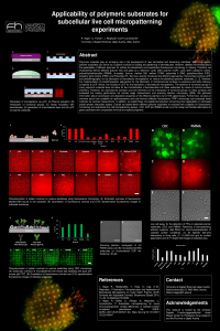
Polymeric materials play an emerging role in the development of new biomedical and biosensing interfaces. Within this regard, polymer substrates can serve as a superior surface for binding and patterning of biomolecules. However, detailed information about the applicability of different polymers for surface functionalization and quantitative fluorescence microscopy is missing. Therefore, we characterized eleven different polymer foils and glass as a reference: cyclic olefin polymer (COP), cyclic olefin copolymer (COC), polymethylmethacrylate (PMMA), di-acetate, lumirror, melinex 506, melinex ST504, polyamide 6 (PA6), polyethersulfone (PES), polyether ether ketone (PEEK) and Polyimide (PI). We have recently introduced two different approaches (microcontact printing (µCP) and photolithography) for the fabrication of biomolecule micropatterns on various functionalized polymer substrates. [1, 2]. However, the implementation of photolithographic approaches for the fabrication of microstructured surfaces is expensive and labor-intensive compared to µCP. Hence, we focused on µCP for the fabrication of biomolecule micropatterns. The absence of functional groups in many polymeric materials does not allow for the immobilization of biomolecules onto these substrates by means of common surface chemistry. Therefore, we used plasma activation and wet chemistry for the introduction of functional groups on these surfaces and evaluated the coating performance via contact angle measurement and scanning electron microscopy (SEM). We gathered information about transmission and absorption properties of the different polymers via UV-VIS spectroscopy. Furthermore, we give an overview about their suitability for epifluorescence and total internal reflection fluorescence (TIRF) microscopy and evaluated these methods via contrast measurement. In addition, we tested these micropatterned polymers concerning their applicability in cell-based protein-protein interaction assays. Overall, we tested eleven different polymer substrates to evaluate their suitability for fluorescence microscopy and subcellular live cell micropatterning assays. COC, COP and PMMA turned out to be cheap and flexible alternatives to glass substrates with comparable chemical and optical properties.
References
- Hager, R.; Haselgrübler, T.; Haas, S.; Lipp, A.-M.; Weghuber, J. Fabrication, Characterization and Application of Biomolecule Micropatterns on Cyclic Olefin Polymer (COP) Surfaces with Adjustable Contrast. Biosensors (Basel) 2019, 10, doi:10.3390/bios10010003.
- Hager, R.; Müller, U.; Ollinger, N.; Weghuber, J.; Lanzerstorfer, P. Subcellular micropatterning for visual immunoprecipitation reveals differences in cytosolic protein complexes downstream the EGFR. bioRxiv 2021.05.25.445547; doi: https://doi.org/10.1101/2021.05.25.445547
Keywords
micropatterns
microcontact printing (µCP)
biomolecules
polymer substrates
protein-protein interactions
live cell assay
Manuscript
Poster Hager Applicability of polymeric substrates for subcellular live cell micropatterning experiments.pdf
Poster
sciforum-048044 Poster Hager Applicability of polymeric substrates for subcellular live cell micropatterning experiments.pdf
0.1 THz imaging with a monolithic High-Tc superconducting transition-edge detector
Design of a Characterization Environment for a MEMS Ultrasound Sensor under Guided Ultrasonic Wave Excitation