Event submissions
Published
 10.3390/eca2022-12742 (registering DOI)
 This submission belongs to the session S2. Mechanisms of Action & Resistance of the event The 2nd International Electronic Conference on Antibiotics
Published date
15 Jun, 2022
Academic Editor
'%3e%3cpath%20d='M12.6657%2010.9104C12.4185%2010.325%2012.0599%209.79327%2011.6097%209.34482C11.1609%208.89508%2010.6293%208.5365%2010.0442%208.28888C10.0389%208.28626%2010.0337%208.28495%2010.0285%208.28233C10.8446%207.69279%2011.3752%206.73249%2011.3752%205.64904C11.3752%203.8542%209.92103%202.39999%208.1262%202.39999C6.33136%202.39999%204.87715%203.8542%204.87715%205.64904C4.87715%206.73249%205.40774%207.69279%206.22393%208.28364C6.21869%208.28626%206.21345%208.28757%206.20821%208.29019C5.62129%208.5378%205.09463%208.89284%204.64265%209.34613C4.1929%209.79493%203.83432%2010.3266%203.58671%2010.9117C3.34345%2011.4845%203.21226%2012.0987%203.20023%2012.7209C3.19988%2012.7349%203.20233%2012.7488%203.20744%2012.7619C3.21255%2012.7749%203.22022%2012.7867%203.22998%2012.7968C3.23975%2012.8068%203.25142%2012.8147%203.26431%2012.8202C3.2772%2012.8256%203.29105%2012.8284%203.30504%2012.8284H4.09109C4.14874%2012.8284%204.19459%2012.7825%204.1959%2012.7262C4.2221%2011.7148%204.62823%2010.7676%205.34617%2010.0497C6.08899%209.30683%207.0755%208.89808%208.1262%208.89808C9.17689%208.89808%2010.1634%209.30683%2010.9062%2010.0497C11.6242%2010.7676%2012.0303%2011.7148%2012.0565%2012.7262C12.0578%2012.7838%2012.1037%2012.8284%2012.1613%2012.8284H12.9474C12.9613%2012.8284%2012.9752%2012.8256%2012.9881%2012.8202C13.001%2012.8147%2013.0126%2012.8068%2013.0224%2012.7968C13.0322%2012.7867%2013.0398%2012.7749%2013.0449%2012.7619C13.0501%2012.7488%2013.0525%2012.7349%2013.0522%2012.7209C13.0391%2012.0947%2012.9094%2011.4855%2012.6657%2010.9104ZM8.1262%207.9024C7.52486%207.9024%206.9589%207.6679%206.53312%207.24211C6.10733%206.81633%205.87283%206.25037%205.87283%205.64904C5.87283%205.0477%206.10733%204.48174%206.53312%204.05596C6.9589%203.63018%207.52486%203.39567%208.1262%203.39567C8.72753%203.39567%209.29349%203.63018%209.71927%204.05596C10.1451%204.48174%2010.3796%205.0477%2010.3796%205.64904C10.3796%206.25037%2010.1451%206.81633%209.71927%207.24211C9.29349%207.6679%208.72753%207.9024%208.1262%207.9024Z'%20fill='%235D1EE1'/%3e%3c/g%3e%3c/svg%3e) Kristine Wang
Kristine WangCitation
Hiral Patel, Kranti Bhamnote, Devarshi Gajjar, Persister cell formation in clinical isolates of Pseudomonas aeruginosa, in Proceedings of The 2nd International Electronic Conference on Antibiotics, 15 June–30 June 2022, MDPI: Basel, Switzerland, doi: 10.3390/eca2022-12742
Share
Email
Facebook
Twitter
LinkedIn
Persister cell formation in clinical isolates of Pseudomonas aeruginosa
'%3e%3cpath%20d='M12.6647%2010.9104C12.4176%2010.325%2012.0589%209.7933%2011.6088%209.34485C11.16%208.89511%2010.6283%208.53653%2010.0432%208.28891C10.038%208.28629%2010.0327%208.28498%2010.0275%208.28236C10.8437%207.69282%2011.3743%206.73252%2011.3743%205.64907C11.3743%203.85423%209.92005%202.40002%208.12522%202.40002C6.33038%202.40002%204.87618%203.85423%204.87618%205.64907C4.87618%206.73252%205.40677%207.69282%206.22296%208.28367C6.21772%208.28629%206.21248%208.2876%206.20724%208.29022C5.62031%208.53783%205.09365%208.89287%204.64167%209.34616C4.19192%209.79496%203.83334%2010.3266%203.58573%2010.9117C3.34247%2011.4846%203.21128%2012.0987%203.19925%2012.721C3.1989%2012.735%203.20135%2012.7489%203.20646%2012.7619C3.21157%2012.7749%203.21924%2012.7868%203.22901%2012.7968C3.23877%2012.8068%203.25045%2012.8148%203.26334%2012.8202C3.27623%2012.8256%203.29007%2012.8284%203.30406%2012.8284H4.09012C4.14776%2012.8284%204.19362%2012.7825%204.19493%2012.7262C4.22113%2011.7148%204.62726%2010.7676%205.34519%2010.0497C6.08802%209.30686%207.07452%208.89811%208.12522%208.89811C9.17592%208.89811%2010.1624%209.30686%2010.9052%2010.0497C11.6232%2010.7676%2012.0293%2011.7148%2012.0555%2012.7262C12.0568%2012.7839%2012.1027%2012.8284%2012.1603%2012.8284H12.9464C12.9604%2012.8284%2012.9742%2012.8256%2012.9871%2012.8202C13%2012.8148%2013.0117%2012.8068%2013.0214%2012.7968C13.0312%2012.7868%2013.0389%2012.7749%2013.044%2012.7619C13.0491%2012.7489%2013.0515%2012.735%2013.0512%2012.721C13.0381%2012.0947%2012.9084%2011.4856%2012.6647%2010.9104ZM8.12522%207.90243C7.52388%207.90243%206.95792%207.66793%206.53214%207.24215C6.10636%206.81636%205.87185%206.2504%205.87185%205.64907C5.87185%205.04773%206.10636%204.48177%206.53214%204.05599C6.95792%203.63021%207.52388%203.3957%208.12522%203.3957C8.72655%203.3957%209.29252%203.63021%209.7183%204.05599C10.1441%204.48177%2010.3786%205.04773%2010.3786%205.64907C10.3786%206.2504%2010.1441%206.81636%209.7183%207.24215C9.29252%207.66793%208.72655%207.90243%208.12522%207.90243Z'%20fill='%235D1EE1'/%3e%3c/g%3e%3c/svg%3e)
Hiral Patel 1
Kranti Bhamnote 1
Devarshi Gajjar 1
1. The Department of Microbiology and Biotechnology Centre, The Maharaja Sayajirao University of Baroda, Vadodara (pin code 390002), Gujarat, India.
Abstract
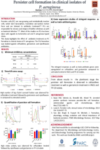
Persister cells (PC) are non-growing and metabolically inactive cells, which lack transcription, translation, and proton motive force. PC formation was reported to occur by (ⅰ) stochastically (ⅱ) antibiotic treatment (ⅲ) nutrient deprivation and (ⅳ) other biofilm-related condition. PCs are responsible for chronic and relapse of biofilm infections as well as bacterial infections. Pseudomonas aeruginosa (Pa) is an opportunistic pathogen frequently causing chronic airway infections in patients with cystic fibrosis (CF), chronic urinary tract infection (UTI), and ventilator-associated pneumonia. For this study, Pa isolates; TP-10, ST-13, and POA1 were used. The Minimum Inhibitory Concentration (MIC) was determined and time-kill assay was carried out for PC isolation in the planktonic stage. Further, Flow Cytometry (FC) and qRT-PCR for stringent response genes (rel A, spo T and lon) and toxin-antitoxin (hig B and hig A) were done. A biphasic kill curve pattern was observed which varied across the antibiotic treatment. Starting from 12-13 log10 cfu/ml mean inoculum, after ceftazidime treatment 9-10 log10 cfu/ml lasted, while on gentamicin and ciprofloxacin treatment 5-6 log10 cfu/ml lasted and formed biphasic pattern. The PC were analyzed through FC using Redox sensor green (RSG) and propidium iodide (PI) staining. It was observed that ceftazidime treatment has high redox activities followed by ciprofloxacin and gentamicin treatment compared to untreated isolate. Whereas ST-13 isolate had high redox activities on ceftazidime treatment followed by gentamicin and ciprofloxacin. The rel A, spo T, lon, hig B and hig A genes were upregulated on ceftazidime and gentamicin treatment compared to untreated in PAO1 and TP-10 isolates. Whereas in ST-13 isolate, genes were upregulated on gentamicin treatment. From the above study, it can be concluded that gentamicin and ceftazidime lead to PCs formation in P. aeruginosa planktonic stage.
Keywords
Persister cell
stringent response
toxin-antitoxin
Pseudomonas aeruginosa
Manuscript
sciforum_061688-done.pdf
Poster
poster-persister-2022.pdf
In vitro activity of cefiderocol and comparators against multi-drug resistant Acinetobacter baumannii isolates
Using antibiotics scaffolds will warrant novel radiotracers for effective positron emission tomography imaging of infections: triumph or pitfall ?