Event submissions
Published
 10.3390/ECMC2022-13462 (registering DOI)
 This submission belongs to the session S6. Emerging technologies in drug discovery of the event 8th International Electronic Conference on Medicinal Chemistry
Published date
01 Nov, 2022
Academic Editor
'%3e%3cpath%20d='M12.6657%2010.9104C12.4185%2010.325%2012.0599%209.79327%2011.6097%209.34482C11.1609%208.89508%2010.6293%208.5365%2010.0442%208.28888C10.0389%208.28626%2010.0337%208.28495%2010.0285%208.28233C10.8446%207.69279%2011.3752%206.73249%2011.3752%205.64904C11.3752%203.8542%209.92103%202.39999%208.1262%202.39999C6.33136%202.39999%204.87715%203.8542%204.87715%205.64904C4.87715%206.73249%205.40774%207.69279%206.22393%208.28364C6.21869%208.28626%206.21345%208.28757%206.20821%208.29019C5.62129%208.5378%205.09463%208.89284%204.64265%209.34613C4.1929%209.79493%203.83432%2010.3266%203.58671%2010.9117C3.34345%2011.4845%203.21226%2012.0987%203.20023%2012.7209C3.19988%2012.7349%203.20233%2012.7488%203.20744%2012.7619C3.21255%2012.7749%203.22022%2012.7867%203.22998%2012.7968C3.23975%2012.8068%203.25142%2012.8147%203.26431%2012.8202C3.2772%2012.8256%203.29105%2012.8284%203.30504%2012.8284H4.09109C4.14874%2012.8284%204.19459%2012.7825%204.1959%2012.7262C4.2221%2011.7148%204.62823%2010.7676%205.34617%2010.0497C6.08899%209.30683%207.0755%208.89808%208.1262%208.89808C9.17689%208.89808%2010.1634%209.30683%2010.9062%2010.0497C11.6242%2010.7676%2012.0303%2011.7148%2012.0565%2012.7262C12.0578%2012.7838%2012.1037%2012.8284%2012.1613%2012.8284H12.9474C12.9613%2012.8284%2012.9752%2012.8256%2012.9881%2012.8202C13.001%2012.8147%2013.0126%2012.8068%2013.0224%2012.7968C13.0322%2012.7867%2013.0398%2012.7749%2013.0449%2012.7619C13.0501%2012.7488%2013.0525%2012.7349%2013.0522%2012.7209C13.0391%2012.0947%2012.9094%2011.4855%2012.6657%2010.9104ZM8.1262%207.9024C7.52486%207.9024%206.9589%207.6679%206.53312%207.24211C6.10733%206.81633%205.87283%206.25037%205.87283%205.64904C5.87283%205.0477%206.10733%204.48174%206.53312%204.05596C6.9589%203.63018%207.52486%203.39567%208.1262%203.39567C8.72753%203.39567%209.29349%203.63018%209.71927%204.05596C10.1451%204.48174%2010.3796%205.0477%2010.3796%205.64904C10.3796%206.25037%2010.1451%206.81633%209.71927%207.24211C9.29349%207.6679%208.72753%207.9024%208.1262%207.9024Z'%20fill='%235D1EE1'/%3e%3c/g%3e%3c/svg%3e) Maria Emília Sousa
Maria Emília SousaCitation
Alexander Dombovski, Ruben Steigerwald, Carina Dilkaute, Bernhard Wünsch, Joachim Jose, Autodisplay of the NMDA receptor GluN1 and GluN2A ligand binding domains as a platform for binding assays, in Proceedings of 8th International Electronic Conference on Medicinal Chemistry, 1 November–30 November 2022, MDPI: Basel, Switzerland, doi: 10.3390/ECMC2022-13462
Share
Email
Facebook
Twitter
LinkedIn
Autodisplay of the NMDA receptor GluN1 and GluN2A ligand binding domains as a platform for binding assays
'%3e%3cpath%20d='M12.6647%2010.9104C12.4176%2010.325%2012.0589%209.7933%2011.6088%209.34485C11.16%208.89511%2010.6283%208.53653%2010.0432%208.28891C10.038%208.28629%2010.0327%208.28498%2010.0275%208.28236C10.8437%207.69282%2011.3743%206.73252%2011.3743%205.64907C11.3743%203.85423%209.92005%202.40002%208.12522%202.40002C6.33038%202.40002%204.87618%203.85423%204.87618%205.64907C4.87618%206.73252%205.40677%207.69282%206.22296%208.28367C6.21772%208.28629%206.21248%208.2876%206.20724%208.29022C5.62031%208.53783%205.09365%208.89287%204.64167%209.34616C4.19192%209.79496%203.83334%2010.3266%203.58573%2010.9117C3.34247%2011.4846%203.21128%2012.0987%203.19925%2012.721C3.1989%2012.735%203.20135%2012.7489%203.20646%2012.7619C3.21157%2012.7749%203.21924%2012.7868%203.22901%2012.7968C3.23877%2012.8068%203.25045%2012.8148%203.26334%2012.8202C3.27623%2012.8256%203.29007%2012.8284%203.30406%2012.8284H4.09012C4.14776%2012.8284%204.19362%2012.7825%204.19493%2012.7262C4.22113%2011.7148%204.62726%2010.7676%205.34519%2010.0497C6.08802%209.30686%207.07452%208.89811%208.12522%208.89811C9.17592%208.89811%2010.1624%209.30686%2010.9052%2010.0497C11.6232%2010.7676%2012.0293%2011.7148%2012.0555%2012.7262C12.0568%2012.7839%2012.1027%2012.8284%2012.1603%2012.8284H12.9464C12.9604%2012.8284%2012.9742%2012.8256%2012.9871%2012.8202C13%2012.8148%2013.0117%2012.8068%2013.0214%2012.7968C13.0312%2012.7868%2013.0389%2012.7749%2013.044%2012.7619C13.0491%2012.7489%2013.0515%2012.735%2013.0512%2012.721C13.0381%2012.0947%2012.9084%2011.4856%2012.6647%2010.9104ZM8.12522%207.90243C7.52388%207.90243%206.95792%207.66793%206.53214%207.24215C6.10636%206.81636%205.87185%206.2504%205.87185%205.64907C5.87185%205.04773%206.10636%204.48177%206.53214%204.05599C6.95792%203.63021%207.52388%203.3957%208.12522%203.3957C8.72655%203.3957%209.29252%203.63021%209.7183%204.05599C10.1441%204.48177%2010.3786%205.04773%2010.3786%205.64907C10.3786%206.2504%2010.1441%206.81636%209.7183%207.24215C9.29252%207.66793%208.72655%207.90243%208.12522%207.90243Z'%20fill='%235D1EE1'/%3e%3c/g%3e%3c/svg%3e)
Alexander Dombovski 1
Ruben Steigerwald 1
Carina Dilkaute 1
Bernhard Wünsch 1

Joachim Jose 1
1. University of Münster, Institute of Pharmaceutical and Medicinal Chemistry, Pharmacampus, Corrensstraße 48, D-48149 Münster, Germany
Abstract
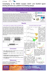
N-methyl-D-aspartate receptors (NMDAR) are heterotetrameric, ionotropic glutamate receptors, typically consisting of two GluN1 and two GluN2 subunits and play a crucial role in neuroplasticity, memory and learning. Seven different subunits lead to a variety of possible subunit combinations in NMDARs. Thus, subunit selective modulation is required for the characterization of properties and (patho)physiological relevance of NMDARs containing different subunits. TCN-201 was the first reported negative allosteric modulator with pronounced selectivity towards GluN2A containing NMDARs. The compound binds at the level of the ligand binding domains (LBD) of the receptor, more precisely at the interface of a local heterodimer of GluN1 and GluN2A LBDs. Three fluorescently labeled ligands based on the scaffold of TCN-201 were developed and autodisplay used as a tool to display the GluN1 and GluN2A LBDs on the surface of E. coli. Surface display was confirmed by proteinase K accessibility assay for GluN1 and GluN2A LBDs individually. Additionally, co-display of both LBDs within one cell sample was established. Ligand binding to surface displayed LBDs was investigated by flow cytometry. Fluorescently labeled ligands were shown to bind to LBD co-displaying cells in a concentration dependant manner. Comparing detection efficiencies suggested that fluorescein-labeled compound 3 was the most suitable tracer molecule. For compound 3 it was additionally shown that practically no off-target binding was detected for non-displaying cells and cells displaying an unrelated dimeric protein. This approach paves the road for developing a competitive binding assay with non-fluorescent ligands to identify novel GluN2A selective high-affinity binders.
Keywords
autodisplay
bacteria
flow cytometry
fluorescent ligand
allosteric modulator
NMDA receptor
Poster
sciforum-066632.pdf
Toxicity evaluation of single and combined exposures to polyhydroxybutyrate nanoparticles and caffeine using Xenopus laevis in vivo and in vitro models
Drug repurposing as an alternative in prostate cancer treatment