Event submissions
Published

This submission belongs to the session A. Biomacromolecules: Proteins of the event The 2nd International Electronic Conference on Biomolecules
Published date
21 Nov, 2022
Academic Editor
'%3e%3cpath%20d='M12.6657%2010.9104C12.4185%2010.325%2012.0599%209.79327%2011.6097%209.34482C11.1609%208.89508%2010.6293%208.5365%2010.0442%208.28888C10.0389%208.28626%2010.0337%208.28495%2010.0285%208.28233C10.8446%207.69279%2011.3752%206.73249%2011.3752%205.64904C11.3752%203.8542%209.92103%202.39999%208.1262%202.39999C6.33136%202.39999%204.87715%203.8542%204.87715%205.64904C4.87715%206.73249%205.40774%207.69279%206.22393%208.28364C6.21869%208.28626%206.21345%208.28757%206.20821%208.29019C5.62129%208.5378%205.09463%208.89284%204.64265%209.34613C4.1929%209.79493%203.83432%2010.3266%203.58671%2010.9117C3.34345%2011.4845%203.21226%2012.0987%203.20023%2012.7209C3.19988%2012.7349%203.20233%2012.7488%203.20744%2012.7619C3.21255%2012.7749%203.22022%2012.7867%203.22998%2012.7968C3.23975%2012.8068%203.25142%2012.8147%203.26431%2012.8202C3.2772%2012.8256%203.29105%2012.8284%203.30504%2012.8284H4.09109C4.14874%2012.8284%204.19459%2012.7825%204.1959%2012.7262C4.2221%2011.7148%204.62823%2010.7676%205.34617%2010.0497C6.08899%209.30683%207.0755%208.89808%208.1262%208.89808C9.17689%208.89808%2010.1634%209.30683%2010.9062%2010.0497C11.6242%2010.7676%2012.0303%2011.7148%2012.0565%2012.7262C12.0578%2012.7838%2012.1037%2012.8284%2012.1613%2012.8284H12.9474C12.9613%2012.8284%2012.9752%2012.8256%2012.9881%2012.8202C13.001%2012.8147%2013.0126%2012.8068%2013.0224%2012.7968C13.0322%2012.7867%2013.0398%2012.7749%2013.0449%2012.7619C13.0501%2012.7488%2013.0525%2012.7349%2013.0522%2012.7209C13.0391%2012.0947%2012.9094%2011.4855%2012.6657%2010.9104ZM8.1262%207.9024C7.52486%207.9024%206.9589%207.6679%206.53312%207.24211C6.10733%206.81633%205.87283%206.25037%205.87283%205.64904C5.87283%205.0477%206.10733%204.48174%206.53312%204.05596C6.9589%203.63018%207.52486%203.39567%208.1262%203.39567C8.72753%203.39567%209.29349%203.63018%209.71927%204.05596C10.1451%204.48174%2010.3796%205.0477%2010.3796%205.64904C10.3796%206.25037%2010.1451%206.81633%209.71927%207.24211C9.29349%207.6679%208.72753%207.9024%208.1262%207.9024Z'%20fill='%235D1EE1'/%3e%3c/g%3e%3c/svg%3e) Cristina Martínez-Villaluenga
Cristina Martínez-VillaluengaCitation
Carlos Marcuello, Anabel Lostao, Recent progress in molecular recognition imaging of protein systems at the nanoscale level, in Proceedings of The 2nd International Electronic Conference on Biomolecules, 1 November–15 November 2022, MDPI: Basel, Switzerland, doi: 10.3390/IECBM2022-13722
Share
Email
Facebook
Twitter
LinkedIn
Recent progress in molecular recognition imaging of protein systems at the nanoscale level

Carlos Marcuello 1,2,3

Anabel Lostao 2,3,4
1. Biosystems and Integrative Sciences (BioISI), Faculdade de Ciências. Universidade de Lisboa 1749-016 Lisboa. Portugal
2. Instituto de Nanociencia y Materiales de Aragón (INMA), CSIC-Universidad de Zaragoza, 50009 Zaragoza. Spain.
3. Laboratorio de Microscopias Avanzadas (LMA), Universidad de Zaragoza, 50018 Zaragoza. Spain.
4. Fundación ARAID, 50018 Zaragoza. Spain.
Abstract
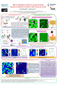
Identification of proteins has received considerable attention in recent years due to the increasing interest to resolve individual biomolecules under physiological relevant conditions. In this framework, atomic force microscopy (AFM), has shown its great potential to acquire a variety of biomolecular physico-chemical properties at the single molecule level [1]. Particularly, force spectroscopy based on AFM (AFM-FS) allows to determine the intermolecular interactions between two biomolecules, requiring being one covalently immobilized on a flat surface and the other linked onto the AFM tip. Previous work was developed in this field by simultaneous topography and recognition imaging (TREC) [2] and tuning-fork-based transverse dynamic force microscopy (TDFM) [3] albeit both methods lack of providing quantitive information. To overcome the aforementioned limitations force-volume (F-V) [4] emerged as promising alternative but the extremely large data acquisition times can lead drifting effects during the image record. Here, we present the intermittent jumping force mode (JM) as suitable approach to gather quantitative high-resolution force maps at local areas of the scanned sample with fast-acquisition times. Using this mode applying very low forces under respulsive regime conditions simultaneous maps of topography and specific rupture forces corresponding to the unbinding of the protein:ligand complexes are obtained. Two different protein systems are employed to illustrate the capabilities of the built up methodological improvements. First, the flavoenzyme system formed between flavodoxin NADP+ reductase (FNR) and its redox partners, ferredoxin and flavodoxin [5, 6] and second, the strongest non-covalent complexes observed in nature between avidin and streptavidin and biotin [7]. In the first case, the results were optimized when an oriented immobilization procedure was designed. In the second work, discrimination between avidin and streptavidin molecules in a hybrid sample was achieved with an unique sensor ligand. The most relevant scientific outcomes can serve as proof-of-principle stage to design diagnostic devices with ultra-sensitivity detection signal for drug screening applications.
[1] Müller, D.J.; Dumitru, A.C.; Lo Giudice, C.; Gaub, H.E.; Hinterdorfer, P.; Hummer, G.; De Yoreo, J.J.; Alsteens, D. Atomic Force Microscopy-Based Force Spectroscopy and Multiparametric Imaging of Biomolecular and Cellular Systems. Chem. Rev. 2020, 121, 11701-11725. https://doi.org/10.1021/acs.chemrev.0c00617.
[2] Stroh, C.M.; Ebner, A.; Geretschlager, M.; Freudenthale, G.; Kienberger, F.; Kamruzzahan, A.S.M.; Smith-Gill, S.J.; Gruber, H.J.; Hinterdorfer, P. Simultaneous topography and recognition imaging using force microscopy. Biophys. J. 2004, 87, 1981-1990. https://doi.org/10.1529/biophysj.104.043331.
[3] Hofer, M.; Adamsmaier, S.; van Zanten, T.S.; Chtcheglova, L.A.; Manzo, C.; Duman, M.; Mayer, B.; Ebner, A.; Moertelmaier, M.; Kada, G.; et al. Molecular recognition imaging using tuning-fork based transverse dynamic force microscopy. Ultramicroscopy 2010, 110, 605-611. https://doi.org/10.1016/j.ultramic.2010.02.019.
[4] Ludwig, M.; Dettmann, W.; Gaub, H.E. Atomic force microscope imaging contrast based on molecular recognition. Biophys. J. 1997, 72, 445-448. https://doi.org/10.1016-3495(97)78685-5.
[5] Marcuello, C.; De Miguel R.; Gómez-Moreno C.; Martínez-Júlvez, M.; Lostao, A. An efficient method for enzyme immobilization evidenced by atomic force microscopy. Protein Eng. Des. Sel. 2012, 25, 715-723. https://doi.org/10.1093/protein/gzs086.
[6] Marcuello, C.; de Miguel, R.; Martínez-Júlvez, M.; Gómez-Moreno, C.; Lostao, A. Mechanostability of the Single-Electron-Transfer Complexes of Anabaena Ferredoxin-NADP+ Reductase. ChemPhysChem. 2015, 16, 3161-3169. https://doi.org/10.1002/cphc.201500534.
[7] Marcuello, C.; De Miguel R.; Lostao, A. Molecular Recognition of Proteins through Quantitative Force Maps at Single Molecule Level. Biomolecules 2022, 12, 594. https://doi.org/10.3390/biom12040594.
Keywords
molecular recognition imaging
atomic force microscopy
functionalized tips
adhesion maps
protein-protein interactions
single molecule level
jumping mode.
Manuscript
sciforum-061472-layout.pdf
Poster
Poster MRI Biomolecules Congress_FINAL.pdf
In silico determination of changes in transcription factor binding sites for the preeclampsia risk haplotype in the regulatory region of the FLT1 gene
Role of APOE Gene Polymorphisms in Coronary Artery Disease