Event submissions
Published
This submission belongs to the session S8. Poster Session of the event The 2nd International Electronic Conference on Microbiology
Published date
26 Dec, 2023
Academic Editor
'%3e%3cpath%20d='M12.6657%2010.9104C12.4185%2010.325%2012.0599%209.79327%2011.6097%209.34482C11.1609%208.89508%2010.6293%208.5365%2010.0442%208.28888C10.0389%208.28626%2010.0337%208.28495%2010.0285%208.28233C10.8446%207.69279%2011.3752%206.73249%2011.3752%205.64904C11.3752%203.8542%209.92103%202.39999%208.1262%202.39999C6.33136%202.39999%204.87715%203.8542%204.87715%205.64904C4.87715%206.73249%205.40774%207.69279%206.22393%208.28364C6.21869%208.28626%206.21345%208.28757%206.20821%208.29019C5.62129%208.5378%205.09463%208.89284%204.64265%209.34613C4.1929%209.79493%203.83432%2010.3266%203.58671%2010.9117C3.34345%2011.4845%203.21226%2012.0987%203.20023%2012.7209C3.19988%2012.7349%203.20233%2012.7488%203.20744%2012.7619C3.21255%2012.7749%203.22022%2012.7867%203.22998%2012.7968C3.23975%2012.8068%203.25142%2012.8147%203.26431%2012.8202C3.2772%2012.8256%203.29105%2012.8284%203.30504%2012.8284H4.09109C4.14874%2012.8284%204.19459%2012.7825%204.1959%2012.7262C4.2221%2011.7148%204.62823%2010.7676%205.34617%2010.0497C6.08899%209.30683%207.0755%208.89808%208.1262%208.89808C9.17689%208.89808%2010.1634%209.30683%2010.9062%2010.0497C11.6242%2010.7676%2012.0303%2011.7148%2012.0565%2012.7262C12.0578%2012.7838%2012.1037%2012.8284%2012.1613%2012.8284H12.9474C12.9613%2012.8284%2012.9752%2012.8256%2012.9881%2012.8202C13.001%2012.8147%2013.0126%2012.8068%2013.0224%2012.7968C13.0322%2012.7867%2013.0398%2012.7749%2013.0449%2012.7619C13.0501%2012.7488%2013.0525%2012.7349%2013.0522%2012.7209C13.0391%2012.0947%2012.9094%2011.4855%2012.6657%2010.9104ZM8.1262%207.9024C7.52486%207.9024%206.9589%207.6679%206.53312%207.24211C6.10733%206.81633%205.87283%206.25037%205.87283%205.64904C5.87283%205.0477%206.10733%204.48174%206.53312%204.05596C6.9589%203.63018%207.52486%203.39567%208.1262%203.39567C8.72753%203.39567%209.29349%203.63018%209.71927%204.05596C10.1451%204.48174%2010.3796%205.0477%2010.3796%205.64904C10.3796%206.25037%2010.1451%206.81633%209.71927%207.24211C9.29349%207.6679%208.72753%207.9024%208.1262%207.9024Z'%20fill='%235D1EE1'/%3e%3c/g%3e%3c/svg%3e) Nico Jehmlich
Nico JehmlichCitation
Giovanna Schiuma, Silvia Beltrami, Sabrina Rizzo, Roberta Rizzo, Angelina Passaro, Pantaleo Greco, Roberta Gafà, Sandra Skuja, Daria Bortolotti, Valerija Groma, GESTATIONAL COVID19: MORPHOLOGICAL ALTERATIONS AND DECREASED HLA-G EXPRESSION CAUSED BY SARS-COV-2 INFECTION, in Proceedings of The 2nd International Electronic Conference on Microbiology, 1 December–15 December 2023, MDPI: Basel, Switzerland
Share
Email
Facebook
Twitter
LinkedIn
GESTATIONAL COVID19: MORPHOLOGICAL ALTERATIONS AND DECREASED HLA-G EXPRESSION CAUSED BY SARS-COV-2 INFECTION
'%3e%3cpath%20d='M12.6647%2010.9104C12.4176%2010.325%2012.0589%209.7933%2011.6088%209.34485C11.16%208.89511%2010.6283%208.53653%2010.0432%208.28891C10.038%208.28629%2010.0327%208.28498%2010.0275%208.28236C10.8437%207.69282%2011.3743%206.73252%2011.3743%205.64907C11.3743%203.85423%209.92005%202.40002%208.12522%202.40002C6.33038%202.40002%204.87618%203.85423%204.87618%205.64907C4.87618%206.73252%205.40677%207.69282%206.22296%208.28367C6.21772%208.28629%206.21248%208.2876%206.20724%208.29022C5.62031%208.53783%205.09365%208.89287%204.64167%209.34616C4.19192%209.79496%203.83334%2010.3266%203.58573%2010.9117C3.34247%2011.4846%203.21128%2012.0987%203.19925%2012.721C3.1989%2012.735%203.20135%2012.7489%203.20646%2012.7619C3.21157%2012.7749%203.21924%2012.7868%203.22901%2012.7968C3.23877%2012.8068%203.25045%2012.8148%203.26334%2012.8202C3.27623%2012.8256%203.29007%2012.8284%203.30406%2012.8284H4.09012C4.14776%2012.8284%204.19362%2012.7825%204.19493%2012.7262C4.22113%2011.7148%204.62726%2010.7676%205.34519%2010.0497C6.08802%209.30686%207.07452%208.89811%208.12522%208.89811C9.17592%208.89811%2010.1624%209.30686%2010.9052%2010.0497C11.6232%2010.7676%2012.0293%2011.7148%2012.0555%2012.7262C12.0568%2012.7839%2012.1027%2012.8284%2012.1603%2012.8284H12.9464C12.9604%2012.8284%2012.9742%2012.8256%2012.9871%2012.8202C13%2012.8148%2013.0117%2012.8068%2013.0214%2012.7968C13.0312%2012.7868%2013.0389%2012.7749%2013.044%2012.7619C13.0491%2012.7489%2013.0515%2012.735%2013.0512%2012.721C13.0381%2012.0947%2012.9084%2011.4856%2012.6647%2010.9104ZM8.12522%207.90243C7.52388%207.90243%206.95792%207.66793%206.53214%207.24215C6.10636%206.81636%205.87185%206.2504%205.87185%205.64907C5.87185%205.04773%206.10636%204.48177%206.53214%204.05599C6.95792%203.63021%207.52388%203.3957%208.12522%203.3957C8.72655%203.3957%209.29252%203.63021%209.7183%204.05599C10.1441%204.48177%2010.3786%205.04773%2010.3786%205.64907C10.3786%206.2504%2010.1441%206.81636%209.7183%207.24215C9.29252%207.66793%208.72655%207.90243%208.12522%207.90243Z'%20fill='%235D1EE1'/%3e%3c/g%3e%3c/svg%3e)
Giovanna Schiuma 1
Silvia Beltrami 1
Sabrina Rizzo 1

Roberta Rizzo 1

Angelina Passaro 1
Pantaleo Greco 1
Roberta Gafà 1
Sandra Skuja 2

Daria Bortolotti 1
Valerija Groma 2
1. University of Ferrara
2. Riga Stradiņš University
Abstract
Background: The evaluation of the effect of SARS-CoV-2 infection during pregnancy has raised interest. Even if virus vertical transmission is still controversial, several researches have focused on the possible distinctive markers associated with different susceptibility to SARS-CoV-2 infection during pregnancy (Greco, Sanz et al. 2023, Schiuma, Beltrami et al. 2023).
Aim: Evaluate the effect of SARS-CoV-2 infection at tissue level in gestational COVID-19
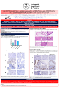
Methods: Morphological alterations were assessed in the placental / chorionic villi, chorionic plate, basal plate, and umbilical cord tissues obtained from 7 subjects with symptomatic respiratory SARS-CoV-2 infection and compared with those in 7 non-COVID control subjects. The expression of SARS-CoV-2 Nucleoprotein (NP) and Human Leukocyte Antigen-G (HLA-G) was estimated by the use of immunohistochemistry.
Results: The 57%, 42,8%, and 28,6% of placental / chorionic villi, chorionic plate, and basal plate, respectively, were found positive for NP antigen (p<0.01), while none of the umbilical cords stained for NP. Placental / chorionic villi samples showed the highest positivity for NP. The presence of NP positivity correlated with high levels of the fibrinoid component in placental / chorionic villi samples and leukocyte infiltration in basal plate. All placental / chorionic villi samples were found positive for HLA-G, independently from NP staining. All the NP positive chorionic plate and half of the NP positive basal plate samples expressed HLA-G. On the contrary, the placental / chorionic villi, chorionic plate, and basal plate of all non-COVID subjects were positive for HLA-G, with a higher H-score in comparison to pathological samples (p<0.05).
Conclusions: The presence of SARS-CoV-2 NP expression in gestational tissues correlates with morphological alterations and a decreased HLA-G expression compared to the control group. These data suggest a possible implication of SARS-CoV-2 infection in morphological and protein expression modification during pregnancy, which might impact infection susceptibility, pregnancy complications, and vertical transmission.
Keywords
COVID-19
pregnancy
HLA-G
immunohistochemistry
Manuscript
Abstract Schiuma NEW'-v2.pdf
Poster
poster Schiuma 2023.pdf
Antimicrobial resistance of foodborne pathogens in pork sausages – A review
Isolation and characterization of agricultural soil bacteria with biotechnological and biological control potential applications