Event submissions
Published
This submission belongs to the session C. Toxicological Mechanisms of Toxicants of the event The 1st International Electronic Conference on Toxics
Published date
03 Apr, 2024
Academic Editor
'%3e%3cpath%20d='M12.6657%2010.9104C12.4185%2010.325%2012.0599%209.79327%2011.6097%209.34482C11.1609%208.89508%2010.6293%208.5365%2010.0442%208.28888C10.0389%208.28626%2010.0337%208.28495%2010.0285%208.28233C10.8446%207.69279%2011.3752%206.73249%2011.3752%205.64904C11.3752%203.8542%209.92103%202.39999%208.1262%202.39999C6.33136%202.39999%204.87715%203.8542%204.87715%205.64904C4.87715%206.73249%205.40774%207.69279%206.22393%208.28364C6.21869%208.28626%206.21345%208.28757%206.20821%208.29019C5.62129%208.5378%205.09463%208.89284%204.64265%209.34613C4.1929%209.79493%203.83432%2010.3266%203.58671%2010.9117C3.34345%2011.4845%203.21226%2012.0987%203.20023%2012.7209C3.19988%2012.7349%203.20233%2012.7488%203.20744%2012.7619C3.21255%2012.7749%203.22022%2012.7867%203.22998%2012.7968C3.23975%2012.8068%203.25142%2012.8147%203.26431%2012.8202C3.2772%2012.8256%203.29105%2012.8284%203.30504%2012.8284H4.09109C4.14874%2012.8284%204.19459%2012.7825%204.1959%2012.7262C4.2221%2011.7148%204.62823%2010.7676%205.34617%2010.0497C6.08899%209.30683%207.0755%208.89808%208.1262%208.89808C9.17689%208.89808%2010.1634%209.30683%2010.9062%2010.0497C11.6242%2010.7676%2012.0303%2011.7148%2012.0565%2012.7262C12.0578%2012.7838%2012.1037%2012.8284%2012.1613%2012.8284H12.9474C12.9613%2012.8284%2012.9752%2012.8256%2012.9881%2012.8202C13.001%2012.8147%2013.0126%2012.8068%2013.0224%2012.7968C13.0322%2012.7867%2013.0398%2012.7749%2013.0449%2012.7619C13.0501%2012.7488%2013.0525%2012.7349%2013.0522%2012.7209C13.0391%2012.0947%2012.9094%2011.4855%2012.6657%2010.9104ZM8.1262%207.9024C7.52486%207.9024%206.9589%207.6679%206.53312%207.24211C6.10733%206.81633%205.87283%206.25037%205.87283%205.64904C5.87283%205.0477%206.10733%204.48174%206.53312%204.05596C6.9589%203.63018%207.52486%203.39567%208.1262%203.39567C8.72753%203.39567%209.29349%203.63018%209.71927%204.05596C10.1451%204.48174%2010.3796%205.0477%2010.3796%205.64904C10.3796%206.25037%2010.1451%206.81633%209.71927%207.24211C9.29349%207.6679%208.72753%207.9024%208.1262%207.9024Z'%20fill='%235D1EE1'/%3e%3c/g%3e%3c/svg%3e) Demetrio Raldúa
Demetrio RaldúaCitation
Jing Hu, The role of the HIF-1α-BNIP3 pathway in acrylonitrile-induced hippocampal neuronal cell toxicity, in Proceedings of The 1st International Electronic Conference on Toxics, 20 March–22 March 2024, MDPI: Basel, Switzerland
Share
Email
Facebook
Twitter
LinkedIn
The role of the HIF-1α-BNIP3 pathway in acrylonitrile-induced hippocampal neuronal cell toxicity
'%3e%3cpath%20d='M12.6647%2010.9104C12.4176%2010.325%2012.0589%209.7933%2011.6088%209.34485C11.16%208.89511%2010.6283%208.53653%2010.0432%208.28891C10.038%208.28629%2010.0327%208.28498%2010.0275%208.28236C10.8437%207.69282%2011.3743%206.73252%2011.3743%205.64907C11.3743%203.85423%209.92005%202.40002%208.12522%202.40002C6.33038%202.40002%204.87618%203.85423%204.87618%205.64907C4.87618%206.73252%205.40677%207.69282%206.22296%208.28367C6.21772%208.28629%206.21248%208.2876%206.20724%208.29022C5.62031%208.53783%205.09365%208.89287%204.64167%209.34616C4.19192%209.79496%203.83334%2010.3266%203.58573%2010.9117C3.34247%2011.4846%203.21128%2012.0987%203.19925%2012.721C3.1989%2012.735%203.20135%2012.7489%203.20646%2012.7619C3.21157%2012.7749%203.21924%2012.7868%203.22901%2012.7968C3.23877%2012.8068%203.25045%2012.8148%203.26334%2012.8202C3.27623%2012.8256%203.29007%2012.8284%203.30406%2012.8284H4.09012C4.14776%2012.8284%204.19362%2012.7825%204.19493%2012.7262C4.22113%2011.7148%204.62726%2010.7676%205.34519%2010.0497C6.08802%209.30686%207.07452%208.89811%208.12522%208.89811C9.17592%208.89811%2010.1624%209.30686%2010.9052%2010.0497C11.6232%2010.7676%2012.0293%2011.7148%2012.0555%2012.7262C12.0568%2012.7839%2012.1027%2012.8284%2012.1603%2012.8284H12.9464C12.9604%2012.8284%2012.9742%2012.8256%2012.9871%2012.8202C13%2012.8148%2013.0117%2012.8068%2013.0214%2012.7968C13.0312%2012.7868%2013.0389%2012.7749%2013.044%2012.7619C13.0491%2012.7489%2013.0515%2012.735%2013.0512%2012.721C13.0381%2012.0947%2012.9084%2011.4856%2012.6647%2010.9104ZM8.12522%207.90243C7.52388%207.90243%206.95792%207.66793%206.53214%207.24215C6.10636%206.81636%205.87185%206.2504%205.87185%205.64907C5.87185%205.04773%206.10636%204.48177%206.53214%204.05599C6.95792%203.63021%207.52388%203.3957%208.12522%203.3957C8.72655%203.3957%209.29252%203.63021%209.7183%204.05599C10.1441%204.48177%2010.3786%205.04773%2010.3786%205.64907C10.3786%206.2504%2010.1441%206.81636%209.7183%207.24215C9.29252%207.66793%208.72655%207.90243%208.12522%207.90243Z'%20fill='%235D1EE1'/%3e%3c/g%3e%3c/svg%3e)
Jing Hu 1
1. Jiangsu Univeisity
Abstract
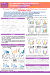
Introduction: Acrylonitrile (AN) is an important chemical raw material, and it is widely used in the production of fibers, plastics and resins. Exposure to AN occurs in occupational settings. While exposure to AN can cause neurotoxicity in bioorganisms, the exact mechanism remains unclear. Hypoxia-inducible factor (HIF) is an important transcription factor produced under hypoxia. During hypoxia, cytoplasm HIF-1α binds to stably expressed HIF-1β and is then translocated into the nucleus to bind with the hypoxia-inducible element (HRE). Multiple downstream target genes are induced.
Methods: CCK assay, LDH release rate, flow cytometry, and Western blot were used to detect the cell viability, toxicity, apoptosis and expression of related proteins in hippocampal neuronal HT22 cells after exposure to AN. Then, cells were pretreated with HIF-1α activator cobalt chloride (CoCl2) and BNIP3 overexpression, followed by the detection of relevant indicators.
Results: We found that the viability of HT22 cells treated with various concentrations of AN (0, 1, 2.5, 5mM) was reduced, but LDH release rate, apoptosis, and ROS were significantly enhanced. The results of Western blotting showed that the expression of HIF-1α and its downstream proteins, including glucose transporter 1, erythropoietin, Bnip3 and NIX were decreased. In addition, due to HIF-1α CoCl2 pretreatment, cell viability and expression of HIF-1α and downstream proteins were increased. CoCl2 increased the expression of BNIP3, TOM20 and BCL2 and decreased the expression of the pro-apoptosis protein BAX. However, HIF-1α inhibitor dimethoxyestradiol (2MEOE2) showed opposite results. Furthermore, we found that cell viability was significantly increased after pretreatment, with BNIP3 overexpression, increased ATP content, and co-localization of BNIP3 and LC3 seen in confocal microscopy. While the expression of TOM20 decreased, the expression of LC3B and Beclin1 increased significantly, indicating that BNIP3 mitophagy may be involved in the process.
Conclusion: The HIF-1α-BNIP3 pathway may participate in the neurotoxic mechanism of AN, providing an effective treatment strategy for clinical AN poisoning.
Keywords
acrylonitrile
neurotoxicity
HT22
HIF-1α;BNIP3;mitophagy
Poster
Jing Hu poster.pdf
Exploring the Role and Regulation of HIF-1 in Manganese Toxicity Using Caenorhabditis
elegans as a Model
elegans as a Model
Characterization of rainbow trout hepatic 3D spheroids for next-generation ecotoxicity testing