Event submissions
Published
This submission belongs to the session F. Biomaterials for Diagnostics, Therapy and Healthcare of the event The 1st International Online Conference on Functional Biomaterials
Published date
08 Jul, 2024
Academic Editor
'%3e%3cpath%20d='M12.6657%2010.9104C12.4185%2010.325%2012.0599%209.79327%2011.6097%209.34482C11.1609%208.89508%2010.6293%208.5365%2010.0442%208.28888C10.0389%208.28626%2010.0337%208.28495%2010.0285%208.28233C10.8446%207.69279%2011.3752%206.73249%2011.3752%205.64904C11.3752%203.8542%209.92103%202.39999%208.1262%202.39999C6.33136%202.39999%204.87715%203.8542%204.87715%205.64904C4.87715%206.73249%205.40774%207.69279%206.22393%208.28364C6.21869%208.28626%206.21345%208.28757%206.20821%208.29019C5.62129%208.5378%205.09463%208.89284%204.64265%209.34613C4.1929%209.79493%203.83432%2010.3266%203.58671%2010.9117C3.34345%2011.4845%203.21226%2012.0987%203.20023%2012.7209C3.19988%2012.7349%203.20233%2012.7488%203.20744%2012.7619C3.21255%2012.7749%203.22022%2012.7867%203.22998%2012.7968C3.23975%2012.8068%203.25142%2012.8147%203.26431%2012.8202C3.2772%2012.8256%203.29105%2012.8284%203.30504%2012.8284H4.09109C4.14874%2012.8284%204.19459%2012.7825%204.1959%2012.7262C4.2221%2011.7148%204.62823%2010.7676%205.34617%2010.0497C6.08899%209.30683%207.0755%208.89808%208.1262%208.89808C9.17689%208.89808%2010.1634%209.30683%2010.9062%2010.0497C11.6242%2010.7676%2012.0303%2011.7148%2012.0565%2012.7262C12.0578%2012.7838%2012.1037%2012.8284%2012.1613%2012.8284H12.9474C12.9613%2012.8284%2012.9752%2012.8256%2012.9881%2012.8202C13.001%2012.8147%2013.0126%2012.8068%2013.0224%2012.7968C13.0322%2012.7867%2013.0398%2012.7749%2013.0449%2012.7619C13.0501%2012.7488%2013.0525%2012.7349%2013.0522%2012.7209C13.0391%2012.0947%2012.9094%2011.4855%2012.6657%2010.9104ZM8.1262%207.9024C7.52486%207.9024%206.9589%207.6679%206.53312%207.24211C6.10733%206.81633%205.87283%206.25037%205.87283%205.64904C5.87283%205.0477%206.10733%204.48174%206.53312%204.05596C6.9589%203.63018%207.52486%203.39567%208.1262%203.39567C8.72753%203.39567%209.29349%203.63018%209.71927%204.05596C10.1451%204.48174%2010.3796%205.0477%2010.3796%205.64904C10.3796%206.25037%2010.1451%206.81633%209.71927%207.24211C9.29349%207.6679%208.72753%207.9024%208.1262%207.9024Z'%20fill='%235D1EE1'/%3e%3c/g%3e%3c/svg%3e) Maria Carmezim
Maria CarmezimCitation
Jainisha Thadhani, G. Roshan Deen, Fryad Henari, Phytosynthesis of Core–Shell Nanoparticles of Selenium and Silver for Biomedical and Environmental Applications, in Proceedings of The 1st International Online Conference on Functional Biomaterials, 10 July–12 July 2024, MDPI: Basel, Switzerland
Share
Email
Facebook
Twitter
LinkedIn
Phytosynthesis of Core–Shell Nanoparticles of Selenium and Silver for Biomedical and Environmental Applications
'%3e%3cpath%20d='M12.6647%2010.9104C12.4176%2010.325%2012.0589%209.7933%2011.6088%209.34485C11.16%208.89511%2010.6283%208.53653%2010.0432%208.28891C10.038%208.28629%2010.0327%208.28498%2010.0275%208.28236C10.8437%207.69282%2011.3743%206.73252%2011.3743%205.64907C11.3743%203.85423%209.92005%202.40002%208.12522%202.40002C6.33038%202.40002%204.87618%203.85423%204.87618%205.64907C4.87618%206.73252%205.40677%207.69282%206.22296%208.28367C6.21772%208.28629%206.21248%208.2876%206.20724%208.29022C5.62031%208.53783%205.09365%208.89287%204.64167%209.34616C4.19192%209.79496%203.83334%2010.3266%203.58573%2010.9117C3.34247%2011.4846%203.21128%2012.0987%203.19925%2012.721C3.1989%2012.735%203.20135%2012.7489%203.20646%2012.7619C3.21157%2012.7749%203.21924%2012.7868%203.22901%2012.7968C3.23877%2012.8068%203.25045%2012.8148%203.26334%2012.8202C3.27623%2012.8256%203.29007%2012.8284%203.30406%2012.8284H4.09012C4.14776%2012.8284%204.19362%2012.7825%204.19493%2012.7262C4.22113%2011.7148%204.62726%2010.7676%205.34519%2010.0497C6.08802%209.30686%207.07452%208.89811%208.12522%208.89811C9.17592%208.89811%2010.1624%209.30686%2010.9052%2010.0497C11.6232%2010.7676%2012.0293%2011.7148%2012.0555%2012.7262C12.0568%2012.7839%2012.1027%2012.8284%2012.1603%2012.8284H12.9464C12.9604%2012.8284%2012.9742%2012.8256%2012.9871%2012.8202C13%2012.8148%2013.0117%2012.8068%2013.0214%2012.7968C13.0312%2012.7868%2013.0389%2012.7749%2013.044%2012.7619C13.0491%2012.7489%2013.0515%2012.735%2013.0512%2012.721C13.0381%2012.0947%2012.9084%2011.4856%2012.6647%2010.9104ZM8.12522%207.90243C7.52388%207.90243%206.95792%207.66793%206.53214%207.24215C6.10636%206.81636%205.87185%206.2504%205.87185%205.64907C5.87185%205.04773%206.10636%204.48177%206.53214%204.05599C6.95792%203.63021%207.52388%203.3957%208.12522%203.3957C8.72655%203.3957%209.29252%203.63021%209.7183%204.05599C10.1441%204.48177%2010.3786%205.04773%2010.3786%205.64907C10.3786%206.2504%2010.1441%206.81636%209.7183%207.24215C9.29252%207.66793%208.72655%207.90243%208.12522%207.90243Z'%20fill='%235D1EE1'/%3e%3c/g%3e%3c/svg%3e)
Jainisha Thadhani 1

G. Roshan Deen 2

Fryad Henari 2
1. School of Medicine, Royal College of Surgeons in Ireland-Medical University of Bahrain, Building No. 2441, Road 2835, Busaiteen Block 228, Muharraq, Busaiteen, 15503, Bahrain
2. Materials for Medicine Research Group, Royal College of Surgeons in Ireland-Medical University of Bahrain, Building No. 2441, Road 2835, Busaiteen Block 228, Muharraq, Busaiteen, 15503, Bahrain
Abstract
1. Introduction
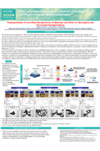
Bimetallic nanoparticles synthesized by non-toxic methods have gained considerable spotlight in applications such as cancer therapy and target-specific drug delivery. This is due to material-specific properties provided by the shell, like metal-specific catalytic and electronic properties and core-associated stability, making them highly customizable. In this project, we successfully synthesized stable bimetallic core–shell particles of silver/selenium for potential applications in drug delivery/antimicrobial use.
2. Methods
Bimetallic nanoparticles of the silver core–selenium shell, and vice versa, were synthesized by a two-step bottom-up approach, using cinnamon, curcumin, and hibiscus plant extracts as part of the green synthesis protocol. Nanoparticles were characterized for size and morphology by UV–Vis spectroscopy, TEM, SEM, and Infra-red spectroscopy.
3. Results
Nanoparticles and raw materials were analysed for nanoparticle formation at each step of
synthesis using UV spectroscopy; the absorption spectra of the extracts were obtained so they could be excluded. Initial plasmon resonance peaks were seen at 285nm in line with the range of 200-350nm expected for selenium, indicating the formation of a selenium core. UV spectrometry was repeated once the silver shell was synthesized, encapsulating the core, and a peak was observed at 446nm in addition to a pre-existing peak at 285nm, inline with expected peaks for silver in the 400-800nm range, indicating the formation of a silver shell. Results were further confirmed with SEM/TEM imaging.
4. Conclusion
Stable bimetallic core–shell nanoparticles were synthesized using plant extracts that acted not only as catalytic compounds but also simultaneous reducing and stabilising agents within the synthesis process, were successfully prepared using inexpensive and eco-friendly methods, as evidenced by UV spectrometry and SEM/TEM imaging showing the presence of spherical nanoparticles measuring under 100nm. Antimicrobial sensitivity testing and further stability testing is currently underway.
Keywords
nanoparticles
green-synthesis
selenium
silver
core-shell
Poster
PHYTO-NP.pdf
ATRP-Synthesized Linear Copolymer Conjugates from Pharmaceutically Functionalized Choline Ionic Liquid Monomers for Ampicillin Delivery
Peptide-mimicking Antifungal Polymers Possessing BBB Penetrating Property to Treat Fungal Infections and Meningitis