Event submissions
Published
This submission belongs to the session S1. Forest Ecology and Management of the event The 4th International Electronic Conference on Forests
Published date
19 Sep, 2024
Academic Editor
'%3e%3cpath%20d='M12.6657%2010.9104C12.4185%2010.325%2012.0599%209.79327%2011.6097%209.34482C11.1609%208.89508%2010.6293%208.5365%2010.0442%208.28888C10.0389%208.28626%2010.0337%208.28495%2010.0285%208.28233C10.8446%207.69279%2011.3752%206.73249%2011.3752%205.64904C11.3752%203.8542%209.92103%202.39999%208.1262%202.39999C6.33136%202.39999%204.87715%203.8542%204.87715%205.64904C4.87715%206.73249%205.40774%207.69279%206.22393%208.28364C6.21869%208.28626%206.21345%208.28757%206.20821%208.29019C5.62129%208.5378%205.09463%208.89284%204.64265%209.34613C4.1929%209.79493%203.83432%2010.3266%203.58671%2010.9117C3.34345%2011.4845%203.21226%2012.0987%203.20023%2012.7209C3.19988%2012.7349%203.20233%2012.7488%203.20744%2012.7619C3.21255%2012.7749%203.22022%2012.7867%203.22998%2012.7968C3.23975%2012.8068%203.25142%2012.8147%203.26431%2012.8202C3.2772%2012.8256%203.29105%2012.8284%203.30504%2012.8284H4.09109C4.14874%2012.8284%204.19459%2012.7825%204.1959%2012.7262C4.2221%2011.7148%204.62823%2010.7676%205.34617%2010.0497C6.08899%209.30683%207.0755%208.89808%208.1262%208.89808C9.17689%208.89808%2010.1634%209.30683%2010.9062%2010.0497C11.6242%2010.7676%2012.0303%2011.7148%2012.0565%2012.7262C12.0578%2012.7838%2012.1037%2012.8284%2012.1613%2012.8284H12.9474C12.9613%2012.8284%2012.9752%2012.8256%2012.9881%2012.8202C13.001%2012.8147%2013.0126%2012.8068%2013.0224%2012.7968C13.0322%2012.7867%2013.0398%2012.7749%2013.0449%2012.7619C13.0501%2012.7488%2013.0525%2012.7349%2013.0522%2012.7209C13.0391%2012.0947%2012.9094%2011.4855%2012.6657%2010.9104ZM8.1262%207.9024C7.52486%207.9024%206.9589%207.6679%206.53312%207.24211C6.10733%206.81633%205.87283%206.25037%205.87283%205.64904C5.87283%205.0477%206.10733%204.48174%206.53312%204.05596C6.9589%203.63018%207.52486%203.39567%208.1262%203.39567C8.72753%203.39567%209.29349%203.63018%209.71927%204.05596C10.1451%204.48174%2010.3796%205.0477%2010.3796%205.64904C10.3796%206.25037%2010.1451%206.81633%209.71927%207.24211C9.29349%207.6679%208.72753%207.9024%208.1262%207.9024Z'%20fill='%235D1EE1'/%3e%3c/g%3e%3c/svg%3e) Giovanna Battipaglia
Giovanna BattipagliaCitation
Timofey Khudykh, Liliana Belokopytova, Bao Yang, Yulia Kholdaenko, Elena Babushkina, Eugene Vaganov, New methods in digital wood anatomy: the automatization of pixel-contrast densitometry and its application across angiosperms, in Proceedings of The 4th International Electronic Conference on Forests, 23 September–25 September 2024, MDPI: Basel, Switzerland
Share
Email
Facebook
Twitter
LinkedIn
New methods in digital wood anatomy: the automatization of pixel-contrast densitometry and its application across angiosperms
'%3e%3cpath%20d='M12.6647%2010.9104C12.4176%2010.325%2012.0589%209.7933%2011.6088%209.34485C11.16%208.89511%2010.6283%208.53653%2010.0432%208.28891C10.038%208.28629%2010.0327%208.28498%2010.0275%208.28236C10.8437%207.69282%2011.3743%206.73252%2011.3743%205.64907C11.3743%203.85423%209.92005%202.40002%208.12522%202.40002C6.33038%202.40002%204.87618%203.85423%204.87618%205.64907C4.87618%206.73252%205.40677%207.69282%206.22296%208.28367C6.21772%208.28629%206.21248%208.2876%206.20724%208.29022C5.62031%208.53783%205.09365%208.89287%204.64167%209.34616C4.19192%209.79496%203.83334%2010.3266%203.58573%2010.9117C3.34247%2011.4846%203.21128%2012.0987%203.19925%2012.721C3.1989%2012.735%203.20135%2012.7489%203.20646%2012.7619C3.21157%2012.7749%203.21924%2012.7868%203.22901%2012.7968C3.23877%2012.8068%203.25045%2012.8148%203.26334%2012.8202C3.27623%2012.8256%203.29007%2012.8284%203.30406%2012.8284H4.09012C4.14776%2012.8284%204.19362%2012.7825%204.19493%2012.7262C4.22113%2011.7148%204.62726%2010.7676%205.34519%2010.0497C6.08802%209.30686%207.07452%208.89811%208.12522%208.89811C9.17592%208.89811%2010.1624%209.30686%2010.9052%2010.0497C11.6232%2010.7676%2012.0293%2011.7148%2012.0555%2012.7262C12.0568%2012.7839%2012.1027%2012.8284%2012.1603%2012.8284H12.9464C12.9604%2012.8284%2012.9742%2012.8256%2012.9871%2012.8202C13%2012.8148%2013.0117%2012.8068%2013.0214%2012.7968C13.0312%2012.7868%2013.0389%2012.7749%2013.044%2012.7619C13.0491%2012.7489%2013.0515%2012.735%2013.0512%2012.721C13.0381%2012.0947%2012.9084%2011.4856%2012.6647%2010.9104ZM8.12522%207.90243C7.52388%207.90243%206.95792%207.66793%206.53214%207.24215C6.10636%206.81636%205.87185%206.2504%205.87185%205.64907C5.87185%205.04773%206.10636%204.48177%206.53214%204.05599C6.95792%203.63021%207.52388%203.3957%208.12522%203.3957C8.72655%203.3957%209.29252%203.63021%209.7183%204.05599C10.1441%204.48177%2010.3786%205.04773%2010.3786%205.64907C10.3786%206.2504%2010.1441%206.81636%209.7183%207.24215C9.29252%207.66793%208.72655%207.90243%208.12522%207.90243Z'%20fill='%235D1EE1'/%3e%3c/g%3e%3c/svg%3e)
Timofey Khudykh 1
Liliana Belokopytova 1,2
Bao Yang 3
Yulia Kholdaenko 1,2
Elena Babushkina 1,2

Eugene Vaganov 1,4
1. Institute of Ecology and Geography, Siberian Federal University, 660041 Krasnoyarsk, Russia
2. Khakass Technical Institute, Siberian Federal University, 655017 Abakan, Russia
3. School of Geography and Ocean Science, Nanjing University, Nanjing 210023, China
4. Department of Dendroecology, V.N. Sukachev Institute of Forest, Siberian Branch of the Russian Academy of
Science, 660036 Krasnoyarsk, Russia
Abstract
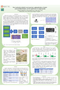
A new method of digital wood anatomy, pixel-contrast densitometry, was proposed recently for conifers. This technique can replace the X-ray densitometer with software that scans and analyzes the image. A photograph of the wood cross-section consists of pixels, i.e., dots of equal area belonging to either the cell wall or the lumen (empty space). By measuring the number of pixels of the cell walls and lumens, the ratio of the cell wall area to the total area of the scan can be determined. The result of multiplying this ratio by the cell wall matter density is the wood density, averaged for the scan area. We used this method to obtain another alternative parameter of wood structure, wood porosity, that is, the proportion of lumen area within scan. We applied this approach to eight angiosperm species of shrubs and trees in Southern Siberia, varying from ring-porous to diffuse-porous wood. The original software was created to implement the technique automatically.
Before analyzing images, their transformations were performed: (1) the Gaussian smoothing algorithm to clean the photo from noise and (2) the Otsu method to separate the cell walls and lumens. Then, a porosity profile over the entire cross-section of the growth ring can be constructed using the program’s virtual sensor.
Good synchronicity between several measurements within ring proved that profiles of wood porosity describe the ring structure and capture inter-annual differences.
For samples of the three most long-living species, Prunus padus L., Caragana arborescens Lam., and Alnus alnobetula Ehrh., 14-32 year-long series of maximum, mean, and minimum porosity determinations and ring widths were analyzed, showing comparable inter-series correlations, common signals, and consistent inter-relations between these parameters of wood. This means that these easily measured characteristics register environmental variability during their formation. Directions for further research were also discussed.
Keywords
digital wood anatomy
densitometry
image analysis
porosity
angiosperms
ring-porous
diffuse-porous
Poster
sciforum-095193_TimofeyKhudykh.pdf
Changes in foliar traits through environmental gradients in two Mediterranean Quercus species and their hybrids.
Modern Genetic and Dynamic Forest Typology: Priority Development Areas and Outstanding Problems