Event submissions
Published
This submission belongs to the session 5. Biosignal Processing of the event The 1st International Online Conference on Bioengineering
Published date
11 Oct, 2024
Academic Editor
'%3e%3cpath%20d='M12.6657%2010.9104C12.4185%2010.325%2012.0599%209.79327%2011.6097%209.34482C11.1609%208.89508%2010.6293%208.5365%2010.0442%208.28888C10.0389%208.28626%2010.0337%208.28495%2010.0285%208.28233C10.8446%207.69279%2011.3752%206.73249%2011.3752%205.64904C11.3752%203.8542%209.92103%202.39999%208.1262%202.39999C6.33136%202.39999%204.87715%203.8542%204.87715%205.64904C4.87715%206.73249%205.40774%207.69279%206.22393%208.28364C6.21869%208.28626%206.21345%208.28757%206.20821%208.29019C5.62129%208.5378%205.09463%208.89284%204.64265%209.34613C4.1929%209.79493%203.83432%2010.3266%203.58671%2010.9117C3.34345%2011.4845%203.21226%2012.0987%203.20023%2012.7209C3.19988%2012.7349%203.20233%2012.7488%203.20744%2012.7619C3.21255%2012.7749%203.22022%2012.7867%203.22998%2012.7968C3.23975%2012.8068%203.25142%2012.8147%203.26431%2012.8202C3.2772%2012.8256%203.29105%2012.8284%203.30504%2012.8284H4.09109C4.14874%2012.8284%204.19459%2012.7825%204.1959%2012.7262C4.2221%2011.7148%204.62823%2010.7676%205.34617%2010.0497C6.08899%209.30683%207.0755%208.89808%208.1262%208.89808C9.17689%208.89808%2010.1634%209.30683%2010.9062%2010.0497C11.6242%2010.7676%2012.0303%2011.7148%2012.0565%2012.7262C12.0578%2012.7838%2012.1037%2012.8284%2012.1613%2012.8284H12.9474C12.9613%2012.8284%2012.9752%2012.8256%2012.9881%2012.8202C13.001%2012.8147%2013.0126%2012.8068%2013.0224%2012.7968C13.0322%2012.7867%2013.0398%2012.7749%2013.0449%2012.7619C13.0501%2012.7488%2013.0525%2012.7349%2013.0522%2012.7209C13.0391%2012.0947%2012.9094%2011.4855%2012.6657%2010.9104ZM8.1262%207.9024C7.52486%207.9024%206.9589%207.6679%206.53312%207.24211C6.10733%206.81633%205.87283%206.25037%205.87283%205.64904C5.87283%205.0477%206.10733%204.48174%206.53312%204.05596C6.9589%203.63018%207.52486%203.39567%208.1262%203.39567C8.72753%203.39567%209.29349%203.63018%209.71927%204.05596C10.1451%204.48174%2010.3796%205.0477%2010.3796%205.64904C10.3796%206.25037%2010.1451%206.81633%209.71927%207.24211C9.29349%207.6679%208.72753%207.9024%208.1262%207.9024Z'%20fill='%235D1EE1'/%3e%3c/g%3e%3c/svg%3e) Andrea Cataldo
Andrea CataldoCitation
DANIELA SOFIA PEDROZO ROCA, JUAN PABLO GRAFFIGNA, VIRGINIA BALLARIN, Functional Characterization of Brain Areas Using Functional Magnetic Resonance Imaging, in Proceedings of The 1st International Online Conference on Bioengineering, 16 October–18 October 2024, MDPI: Basel, Switzerland
Share
Email
Facebook
Twitter
LinkedIn
Functional Characterization of Brain Areas Using Functional Magnetic Resonance Imaging
'%3e%3cpath%20d='M12.6647%2010.9104C12.4176%2010.325%2012.0589%209.7933%2011.6088%209.34485C11.16%208.89511%2010.6283%208.53653%2010.0432%208.28891C10.038%208.28629%2010.0327%208.28498%2010.0275%208.28236C10.8437%207.69282%2011.3743%206.73252%2011.3743%205.64907C11.3743%203.85423%209.92005%202.40002%208.12522%202.40002C6.33038%202.40002%204.87618%203.85423%204.87618%205.64907C4.87618%206.73252%205.40677%207.69282%206.22296%208.28367C6.21772%208.28629%206.21248%208.2876%206.20724%208.29022C5.62031%208.53783%205.09365%208.89287%204.64167%209.34616C4.19192%209.79496%203.83334%2010.3266%203.58573%2010.9117C3.34247%2011.4846%203.21128%2012.0987%203.19925%2012.721C3.1989%2012.735%203.20135%2012.7489%203.20646%2012.7619C3.21157%2012.7749%203.21924%2012.7868%203.22901%2012.7968C3.23877%2012.8068%203.25045%2012.8148%203.26334%2012.8202C3.27623%2012.8256%203.29007%2012.8284%203.30406%2012.8284H4.09012C4.14776%2012.8284%204.19362%2012.7825%204.19493%2012.7262C4.22113%2011.7148%204.62726%2010.7676%205.34519%2010.0497C6.08802%209.30686%207.07452%208.89811%208.12522%208.89811C9.17592%208.89811%2010.1624%209.30686%2010.9052%2010.0497C11.6232%2010.7676%2012.0293%2011.7148%2012.0555%2012.7262C12.0568%2012.7839%2012.1027%2012.8284%2012.1603%2012.8284H12.9464C12.9604%2012.8284%2012.9742%2012.8256%2012.9871%2012.8202C13%2012.8148%2013.0117%2012.8068%2013.0214%2012.7968C13.0312%2012.7868%2013.0389%2012.7749%2013.044%2012.7619C13.0491%2012.7489%2013.0515%2012.735%2013.0512%2012.721C13.0381%2012.0947%2012.9084%2011.4856%2012.6647%2010.9104ZM8.12522%207.90243C7.52388%207.90243%206.95792%207.66793%206.53214%207.24215C6.10636%206.81636%205.87185%206.2504%205.87185%205.64907C5.87185%205.04773%206.10636%204.48177%206.53214%204.05599C6.95792%203.63021%207.52388%203.3957%208.12522%203.3957C8.72655%203.3957%209.29252%203.63021%209.7183%204.05599C10.1441%204.48177%2010.3786%205.04773%2010.3786%205.64907C10.3786%206.2504%2010.1441%206.81636%209.7183%207.24215C9.29252%207.66793%208.72655%207.90243%208.12522%207.90243Z'%20fill='%235D1EE1'/%3e%3c/g%3e%3c/svg%3e)
DANIELA SOFIA PEDROZO ROCA 1
JUAN PABLO GRAFFIGNA 1
VIRGINIA BALLARIN 2
1. INBIO, UNSJ, San Juan, Argentina: Libertador Gral. San Martín (West) 1109, San Juan, Argentina (ZC: J5400ARL)
2. FI, UNMDP, Mar del Plata, Argentina: Juan B. Justo 4302, Mar del Plata, Buenos Aires, Argentina (ZC: B7608FDQ)
Abstract
Introduction
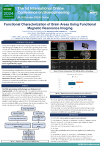
Functional magnetic resonance imaging (fMRI) is a non-invasive neuroimaging modality that is continuously growing, both in the clinical and scientific fields. The analysis of these images requires a very complex and varied post-processing of the obtained images. This causes the results of different studies to be non-comparable or difficult to characterize. In order to simplify the processing and obtain objective results with analyzable metrics, this work proposes the development of an analysis methodology to obtain statistical values on brain activation areas segmented by region.
Methods
Several specialized tools were used. First, the FreeSurfer scientific package was used for brain segmentation. Then, widely used software for the analysis of fMRI data, FSL, was used for activation areas. The last processing and analysis steps were performed with the 3D Slicer medical image visualization tool. Based on these tools, a method was defined to obtain fMRI activation metrics for each of the 148 brain structures obtained by FreeSurfer.
This method was applied to the database proposed by [Masterson TD et al., 2016], where studies of neuronal response to visual food stimuli were carried out at two different times of the day. From this, it is possible to perform a comparative analysis of different metrics for each functional area and to define the levels of significance.
Results
A method was obtained for generating metrics that characterize functional studies based on the use of open-source scientific tools. This method was then applied to a database of fMRI images. Finally, a non-parametric statistical study was used based on the characterization of each area for all subjects, obtaining the most significant features.
Conclusion
This work applied a methodology for automatic fMRI image processing to obtain metrics and perform the most convenient statistical analyses. This method can be extended to different intra- and inter-patient comparisons.
Keywords
fMRI
FSL
FreeSurfer
3D Slicer
Image processing
Poster
poster-sciforum-101179.pdf
SYMMETRY-BASED EYE DETECTION IN FACIAL IMAGES USING HOUGH TRANSFORM FOR CIRCLES
A GC-MS and network pharmacology based approach for investigating anti-inflammatory mechanism of compounds extracted from moss Barbula constricta