Event submissions
Published
This submission belongs to the session 1. Regenerative and Tissue Engineering of the event The 1st International Online Conference on Bioengineering
Published date
11 Oct, 2024
Academic Editor
'%3e%3cpath%20d='M12.6657%2010.9104C12.4185%2010.325%2012.0599%209.79327%2011.6097%209.34482C11.1609%208.89508%2010.6293%208.5365%2010.0442%208.28888C10.0389%208.28626%2010.0337%208.28495%2010.0285%208.28233C10.8446%207.69279%2011.3752%206.73249%2011.3752%205.64904C11.3752%203.8542%209.92103%202.39999%208.1262%202.39999C6.33136%202.39999%204.87715%203.8542%204.87715%205.64904C4.87715%206.73249%205.40774%207.69279%206.22393%208.28364C6.21869%208.28626%206.21345%208.28757%206.20821%208.29019C5.62129%208.5378%205.09463%208.89284%204.64265%209.34613C4.1929%209.79493%203.83432%2010.3266%203.58671%2010.9117C3.34345%2011.4845%203.21226%2012.0987%203.20023%2012.7209C3.19988%2012.7349%203.20233%2012.7488%203.20744%2012.7619C3.21255%2012.7749%203.22022%2012.7867%203.22998%2012.7968C3.23975%2012.8068%203.25142%2012.8147%203.26431%2012.8202C3.2772%2012.8256%203.29105%2012.8284%203.30504%2012.8284H4.09109C4.14874%2012.8284%204.19459%2012.7825%204.1959%2012.7262C4.2221%2011.7148%204.62823%2010.7676%205.34617%2010.0497C6.08899%209.30683%207.0755%208.89808%208.1262%208.89808C9.17689%208.89808%2010.1634%209.30683%2010.9062%2010.0497C11.6242%2010.7676%2012.0303%2011.7148%2012.0565%2012.7262C12.0578%2012.7838%2012.1037%2012.8284%2012.1613%2012.8284H12.9474C12.9613%2012.8284%2012.9752%2012.8256%2012.9881%2012.8202C13.001%2012.8147%2013.0126%2012.8068%2013.0224%2012.7968C13.0322%2012.7867%2013.0398%2012.7749%2013.0449%2012.7619C13.0501%2012.7488%2013.0525%2012.7349%2013.0522%2012.7209C13.0391%2012.0947%2012.9094%2011.4855%2012.6657%2010.9104ZM8.1262%207.9024C7.52486%207.9024%206.9589%207.6679%206.53312%207.24211C6.10733%206.81633%205.87283%206.25037%205.87283%205.64904C5.87283%205.0477%206.10733%204.48174%206.53312%204.05596C6.9589%203.63018%207.52486%203.39567%208.1262%203.39567C8.72753%203.39567%209.29349%203.63018%209.71927%204.05596C10.1451%204.48174%2010.3796%205.0477%2010.3796%205.64904C10.3796%206.25037%2010.1451%206.81633%209.71927%207.24211C9.29349%207.6679%208.72753%207.9024%208.1262%207.9024Z'%20fill='%235D1EE1'/%3e%3c/g%3e%3c/svg%3e) Elena A Jones
Elena A JonesCitation
Débora Czarnabay, Fernanda Muckler, Marcelo Garrido dos Santos, Laura Elena Sperling, Patricia Pranke, Cutting-Edge Bioink Technology: Using Decellularized Skin for Enhanced 3D Bioprinting, in Proceedings of The 1st International Online Conference on Bioengineering, 16 October–18 October 2024, MDPI: Basel, Switzerland
Share
Email
Facebook
Twitter
LinkedIn
Cutting-Edge Bioink Technology: Using Decellularized Skin for Enhanced 3D Bioprinting
'%3e%3cpath%20d='M12.6647%2010.9104C12.4176%2010.325%2012.0589%209.7933%2011.6088%209.34485C11.16%208.89511%2010.6283%208.53653%2010.0432%208.28891C10.038%208.28629%2010.0327%208.28498%2010.0275%208.28236C10.8437%207.69282%2011.3743%206.73252%2011.3743%205.64907C11.3743%203.85423%209.92005%202.40002%208.12522%202.40002C6.33038%202.40002%204.87618%203.85423%204.87618%205.64907C4.87618%206.73252%205.40677%207.69282%206.22296%208.28367C6.21772%208.28629%206.21248%208.2876%206.20724%208.29022C5.62031%208.53783%205.09365%208.89287%204.64167%209.34616C4.19192%209.79496%203.83334%2010.3266%203.58573%2010.9117C3.34247%2011.4846%203.21128%2012.0987%203.19925%2012.721C3.1989%2012.735%203.20135%2012.7489%203.20646%2012.7619C3.21157%2012.7749%203.21924%2012.7868%203.22901%2012.7968C3.23877%2012.8068%203.25045%2012.8148%203.26334%2012.8202C3.27623%2012.8256%203.29007%2012.8284%203.30406%2012.8284H4.09012C4.14776%2012.8284%204.19362%2012.7825%204.19493%2012.7262C4.22113%2011.7148%204.62726%2010.7676%205.34519%2010.0497C6.08802%209.30686%207.07452%208.89811%208.12522%208.89811C9.17592%208.89811%2010.1624%209.30686%2010.9052%2010.0497C11.6232%2010.7676%2012.0293%2011.7148%2012.0555%2012.7262C12.0568%2012.7839%2012.1027%2012.8284%2012.1603%2012.8284H12.9464C12.9604%2012.8284%2012.9742%2012.8256%2012.9871%2012.8202C13%2012.8148%2013.0117%2012.8068%2013.0214%2012.7968C13.0312%2012.7868%2013.0389%2012.7749%2013.044%2012.7619C13.0491%2012.7489%2013.0515%2012.735%2013.0512%2012.721C13.0381%2012.0947%2012.9084%2011.4856%2012.6647%2010.9104ZM8.12522%207.90243C7.52388%207.90243%206.95792%207.66793%206.53214%207.24215C6.10636%206.81636%205.87185%206.2504%205.87185%205.64907C5.87185%205.04773%206.10636%204.48177%206.53214%204.05599C6.95792%203.63021%207.52388%203.3957%208.12522%203.3957C8.72655%203.3957%209.29252%203.63021%209.7183%204.05599C10.1441%204.48177%2010.3786%205.04773%2010.3786%205.64907C10.3786%206.2504%2010.1441%206.81636%209.7183%207.24215C9.29252%207.66793%208.72655%207.90243%208.12522%207.90243Z'%20fill='%235D1EE1'/%3e%3c/g%3e%3c/svg%3e)
Débora Czarnabay 1
Fernanda Muckler 2
Marcelo Garrido dos Santos 3
Laura Elena Sperling 1,4
Patricia Pranke 1,5
1. Stem Cell Research Institute, Universidade Federal do Rio Grande do Sul, Porto Alegre, Rio Grande do Sul, Brazil
2. Universidade Federal do Rio Grande do Sul, Porto Alegre, Rio Grande do Sul, Brazil
3. Department of Phisiology/ Universidade Federal do Rio Grande do Sul, Porto Alegre, Rio Grande do Sul, Brazil
4. Universidade do Vale do Rio dos Sinos, São Leopoldo, Rio Grande do Sul, Brazil
5. Hematology and Stem Cell Laboratory, Faculty of Pharmacy, Universidade Federal do Rio Grande do Sul, Porto Alegre, Rio Grande do Sul, Brazil
Abstract
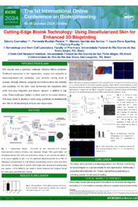
Introduction: Skin wounds present a considerable challenge, impacting millions globally. This research aimed to create a bioink utilizing lyophilized rat decellularized skin (DS) for 3D bioprinting to improve skin regeneration. Methods: Rat skin was subjected to decellularization for 5 days. A comparative analysis of genomic DNA quantification and histological staining was performed between native and decellularized skin. The tissue was freeze-dried and combined with alginate and gelatin to formulate bioinks with concentrations of 1.5% and 3% DS, 3% or 4% alginate, and 7% gelatin. Rheological evaluations, including swelling, printability, and degradation over a four-week period, were conducted. Hydrogel SEM images were obtained using a scanning electron microscope. Cell viability and proliferation were assessed using the Live/Dead assay.
Results: The hydrogel demonstrated good shear-thinning behavior and maintained its viscosity across different concentrations. The degradation rate was 59,2% in one month. Swelling was 3783% after one month. Only the bioink with 1.5% DS, 3% alginate, and 7% gelatin preserved structural integrity for four weeks and was chosen for further examination. Furthermore, the bioink showed a low tangent delta, decreasing printing-related stress and subsequent cell death. SEM images revealed a porous three-dimensional structure. The Live/Dead assay indicated higher cell viability (65%) compared to the control seven days post-bioprinting.
Conclusion: The biomaterial showed good mechanical properties and, after bioprinting, supported cell proliferation, indicating its potential as a promising alternative for skin wound regeneration.
Funding: The Office of Naval Research Global (ONRG Award N62909-21-1-2026), The National Institute of Science and Technology for Regenerative Medicine (INCT-Regenera) and The Stem Cell Research Institute (IPCT).
Keywords
Skin regeneration
wound healing
3D bioprinting
Bioink
decellularization
Poster
Poster IOCBE2024_.pdf
Naturally derived biopolymers in 3D bioprinting for biomedical applications
Transfected Nicotiana tabacum BY-2 cell cultivation in a bioreactor with wave-induced agitation