Event submissions
Published
This submission belongs to the session 1. Regenerative and Tissue Engineering of the event The 1st International Online Conference on Bioengineering
Published date
11 Oct, 2024
Academic Editor
'%3e%3cpath%20d='M12.6657%2010.9104C12.4185%2010.325%2012.0599%209.79327%2011.6097%209.34482C11.1609%208.89508%2010.6293%208.5365%2010.0442%208.28888C10.0389%208.28626%2010.0337%208.28495%2010.0285%208.28233C10.8446%207.69279%2011.3752%206.73249%2011.3752%205.64904C11.3752%203.8542%209.92103%202.39999%208.1262%202.39999C6.33136%202.39999%204.87715%203.8542%204.87715%205.64904C4.87715%206.73249%205.40774%207.69279%206.22393%208.28364C6.21869%208.28626%206.21345%208.28757%206.20821%208.29019C5.62129%208.5378%205.09463%208.89284%204.64265%209.34613C4.1929%209.79493%203.83432%2010.3266%203.58671%2010.9117C3.34345%2011.4845%203.21226%2012.0987%203.20023%2012.7209C3.19988%2012.7349%203.20233%2012.7488%203.20744%2012.7619C3.21255%2012.7749%203.22022%2012.7867%203.22998%2012.7968C3.23975%2012.8068%203.25142%2012.8147%203.26431%2012.8202C3.2772%2012.8256%203.29105%2012.8284%203.30504%2012.8284H4.09109C4.14874%2012.8284%204.19459%2012.7825%204.1959%2012.7262C4.2221%2011.7148%204.62823%2010.7676%205.34617%2010.0497C6.08899%209.30683%207.0755%208.89808%208.1262%208.89808C9.17689%208.89808%2010.1634%209.30683%2010.9062%2010.0497C11.6242%2010.7676%2012.0303%2011.7148%2012.0565%2012.7262C12.0578%2012.7838%2012.1037%2012.8284%2012.1613%2012.8284H12.9474C12.9613%2012.8284%2012.9752%2012.8256%2012.9881%2012.8202C13.001%2012.8147%2013.0126%2012.8068%2013.0224%2012.7968C13.0322%2012.7867%2013.0398%2012.7749%2013.0449%2012.7619C13.0501%2012.7488%2013.0525%2012.7349%2013.0522%2012.7209C13.0391%2012.0947%2012.9094%2011.4855%2012.6657%2010.9104ZM8.1262%207.9024C7.52486%207.9024%206.9589%207.6679%206.53312%207.24211C6.10733%206.81633%205.87283%206.25037%205.87283%205.64904C5.87283%205.0477%206.10733%204.48174%206.53312%204.05596C6.9589%203.63018%207.52486%203.39567%208.1262%203.39567C8.72753%203.39567%209.29349%203.63018%209.71927%204.05596C10.1451%204.48174%2010.3796%205.0477%2010.3796%205.64904C10.3796%206.25037%2010.1451%206.81633%209.71927%207.24211C9.29349%207.6679%208.72753%207.9024%208.1262%207.9024Z'%20fill='%235D1EE1'/%3e%3c/g%3e%3c/svg%3e) Elena A Jones
Elena A JonesCitation
Lucas Eduardo Negreiros, Marcelo Garrido dos Santos, Julia Santos, Fernanda Muckler, Natalia Santos, Debora Czarnabay, Laura Sperling, Patricia Pranke, Three-dimensional-bioprinted bioink with nanosilicate and pluronic p123 for bone tissue engineering, in Proceedings of The 1st International Online Conference on Bioengineering, 16 October–18 October 2024, MDPI: Basel, Switzerland
Share
Email
Facebook
Twitter
LinkedIn
Three-dimensional-bioprinted bioink with nanosilicate and pluronic p123 for bone tissue engineering
'%3e%3cpath%20d='M12.6647%2010.9104C12.4176%2010.325%2012.0589%209.7933%2011.6088%209.34485C11.16%208.89511%2010.6283%208.53653%2010.0432%208.28891C10.038%208.28629%2010.0327%208.28498%2010.0275%208.28236C10.8437%207.69282%2011.3743%206.73252%2011.3743%205.64907C11.3743%203.85423%209.92005%202.40002%208.12522%202.40002C6.33038%202.40002%204.87618%203.85423%204.87618%205.64907C4.87618%206.73252%205.40677%207.69282%206.22296%208.28367C6.21772%208.28629%206.21248%208.2876%206.20724%208.29022C5.62031%208.53783%205.09365%208.89287%204.64167%209.34616C4.19192%209.79496%203.83334%2010.3266%203.58573%2010.9117C3.34247%2011.4846%203.21128%2012.0987%203.19925%2012.721C3.1989%2012.735%203.20135%2012.7489%203.20646%2012.7619C3.21157%2012.7749%203.21924%2012.7868%203.22901%2012.7968C3.23877%2012.8068%203.25045%2012.8148%203.26334%2012.8202C3.27623%2012.8256%203.29007%2012.8284%203.30406%2012.8284H4.09012C4.14776%2012.8284%204.19362%2012.7825%204.19493%2012.7262C4.22113%2011.7148%204.62726%2010.7676%205.34519%2010.0497C6.08802%209.30686%207.07452%208.89811%208.12522%208.89811C9.17592%208.89811%2010.1624%209.30686%2010.9052%2010.0497C11.6232%2010.7676%2012.0293%2011.7148%2012.0555%2012.7262C12.0568%2012.7839%2012.1027%2012.8284%2012.1603%2012.8284H12.9464C12.9604%2012.8284%2012.9742%2012.8256%2012.9871%2012.8202C13%2012.8148%2013.0117%2012.8068%2013.0214%2012.7968C13.0312%2012.7868%2013.0389%2012.7749%2013.044%2012.7619C13.0491%2012.7489%2013.0515%2012.735%2013.0512%2012.721C13.0381%2012.0947%2012.9084%2011.4856%2012.6647%2010.9104ZM8.12522%207.90243C7.52388%207.90243%206.95792%207.66793%206.53214%207.24215C6.10636%206.81636%205.87185%206.2504%205.87185%205.64907C5.87185%205.04773%206.10636%204.48177%206.53214%204.05599C6.95792%203.63021%207.52388%203.3957%208.12522%203.3957C8.72655%203.3957%209.29252%203.63021%209.7183%204.05599C10.1441%204.48177%2010.3786%205.04773%2010.3786%205.64907C10.3786%206.2504%2010.1441%206.81636%209.7183%207.24215C9.29252%207.66793%208.72655%207.90243%208.12522%207.90243Z'%20fill='%235D1EE1'/%3e%3c/g%3e%3c/svg%3e)
Lucas Eduardo Negreiros 1
Marcelo Garrido dos Santos 2
Julia Santos 3
Fernanda Muckler 4
Natalia Santos 5
Debora Czarnabay 4
Laura Sperling 6
Patricia Pranke 4
1. Faculty of Pharmacy, Federal University of Rio Grande do Sul, Porto Alegre - RS, Brazil.
2. Postgraduate program of Physiology, Federal University of Rio Grande do Sul, Porto Alegre - RS, Brazil.
3. Federal University of Health Sciences of Porto Alegre, Porto Alegre - RS, Brazil.
4. Federal University of Rio Grande do Sul, Porto Alegre - RS, Brazil.
5. Chemistry Institute, Federal University of Rio Grande do Sul, Porto Alegre - RS, Brazil.
6. Federal University of Rio Grande do Sul, Porto Alegre - RS, Brazil.
Unisinos, São Leopoldo - RS, Brazil.
Abstract
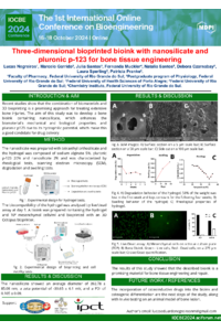
Introduction: Recent studies show that the combination of biomaterials and 3D bioprinting is a promising approach for treating extensive bone injuries. The aim of this study was to develop a bone bioink containing nanosilicate, which enhances the biomaterial's mechanical and biological properties, and pluronic p123 due to its hydrophilic potential, which make this a good candidate for drug delivery. Methodology: Nanosilicate was prepared with tetraethyl orthosilicate (TEOS) and the hydrogel was composed of alginate 5%, pluronic p123 20%, and nanosilicate 2%; it was characterized using rheological tests, scanning electron microscopy (SEM), and degradation and swelling tests. The hydrogel was then mixed with 106 mesenchymal cells/mL and was bioprinted in a 3D bioprinter. In order to evaluate the biocompatibility, a live/dead assay was performed on day one. Results: The nanosilicate showed an average diameter of 392.78 ± 85.08 nm, a zeta potential of -39.65 ± 6.1 mV, and a PDI of 0.105 ± 0.09. The SEM images showed the highly porous structure of the hydrogel with distribution of the nanosilicate throughout the surface of the pores, suggesting an optimal structure for cell adhesion. The hydrogel swelled 1718% over 24 hours, indicating a good capacity for nutrient exchange and cell migration. The degradation rate was 54.21% after one month, suggesting a good clearance of the biomaterial during the regeneration of the natural tissue. Rheological characterization showed a suitable G´/ G´´ ratio, indicating good mechanical properties to biomimic the bone tissue. The live/dead assay revealed a cell viability of around 70% after one day, being non-cytotoxic for the mesenchymal cells. Conclusion: The results of this study showed that the described bioink is a promising material for bone tissue engineering and repair.
Keywords
Bone regeneration
3D Bioprinting
Nanosilicate
Mesenchymal
Poster
097718.pdf
Stress Detection using Bio-Signal Processing: An application of IoT and ML for Old Age Home Residents
Development of antimicrobial PDMS polymers containing Silver-Copper Nanoparticles for potential applications in biomedical devices