Event submissions
Published
This submission belongs to the session S1. Biophotonics and Biomedical Optics of the event The 1st International Online Conference on Photonics
Published date
14 Oct, 2024
Academic Editor
'%3e%3cpath%20d='M12.6657%2010.9104C12.4185%2010.325%2012.0599%209.79327%2011.6097%209.34482C11.1609%208.89508%2010.6293%208.5365%2010.0442%208.28888C10.0389%208.28626%2010.0337%208.28495%2010.0285%208.28233C10.8446%207.69279%2011.3752%206.73249%2011.3752%205.64904C11.3752%203.8542%209.92103%202.39999%208.1262%202.39999C6.33136%202.39999%204.87715%203.8542%204.87715%205.64904C4.87715%206.73249%205.40774%207.69279%206.22393%208.28364C6.21869%208.28626%206.21345%208.28757%206.20821%208.29019C5.62129%208.5378%205.09463%208.89284%204.64265%209.34613C4.1929%209.79493%203.83432%2010.3266%203.58671%2010.9117C3.34345%2011.4845%203.21226%2012.0987%203.20023%2012.7209C3.19988%2012.7349%203.20233%2012.7488%203.20744%2012.7619C3.21255%2012.7749%203.22022%2012.7867%203.22998%2012.7968C3.23975%2012.8068%203.25142%2012.8147%203.26431%2012.8202C3.2772%2012.8256%203.29105%2012.8284%203.30504%2012.8284H4.09109C4.14874%2012.8284%204.19459%2012.7825%204.1959%2012.7262C4.2221%2011.7148%204.62823%2010.7676%205.34617%2010.0497C6.08899%209.30683%207.0755%208.89808%208.1262%208.89808C9.17689%208.89808%2010.1634%209.30683%2010.9062%2010.0497C11.6242%2010.7676%2012.0303%2011.7148%2012.0565%2012.7262C12.0578%2012.7838%2012.1037%2012.8284%2012.1613%2012.8284H12.9474C12.9613%2012.8284%2012.9752%2012.8256%2012.9881%2012.8202C13.001%2012.8147%2013.0126%2012.8068%2013.0224%2012.7968C13.0322%2012.7867%2013.0398%2012.7749%2013.0449%2012.7619C13.0501%2012.7488%2013.0525%2012.7349%2013.0522%2012.7209C13.0391%2012.0947%2012.9094%2011.4855%2012.6657%2010.9104ZM8.1262%207.9024C7.52486%207.9024%206.9589%207.6679%206.53312%207.24211C6.10733%206.81633%205.87283%206.25037%205.87283%205.64904C5.87283%205.0477%206.10733%204.48174%206.53312%204.05596C6.9589%203.63018%207.52486%203.39567%208.1262%203.39567C8.72753%203.39567%209.29349%203.63018%209.71927%204.05596C10.1451%204.48174%2010.3796%205.0477%2010.3796%205.64904C10.3796%206.25037%2010.1451%206.81633%209.71927%207.24211C9.29349%207.6679%208.72753%207.9024%208.1262%207.9024Z'%20fill='%235D1EE1'/%3e%3c/g%3e%3c/svg%3e) Ambra Giannetti
Ambra GiannettiCitation
Nora Dassmann, Bennet Brockhagen, Andrea Ehrmann, Investigation of the optical contrast between nanofiber mats and mammalian cells dyed with fluorescent and other dyes, in Proceedings of The 1st International Online Conference on Photonics, 14 October–16 October 2024, MDPI: Basel, Switzerland
Share
Email
Facebook
Twitter
LinkedIn
Investigation of the optical contrast between nanofiber mats and mammalian cells dyed with fluorescent and other dyes
'%3e%3cpath%20d='M12.6647%2010.9104C12.4176%2010.325%2012.0589%209.7933%2011.6088%209.34485C11.16%208.89511%2010.6283%208.53653%2010.0432%208.28891C10.038%208.28629%2010.0327%208.28498%2010.0275%208.28236C10.8437%207.69282%2011.3743%206.73252%2011.3743%205.64907C11.3743%203.85423%209.92005%202.40002%208.12522%202.40002C6.33038%202.40002%204.87618%203.85423%204.87618%205.64907C4.87618%206.73252%205.40677%207.69282%206.22296%208.28367C6.21772%208.28629%206.21248%208.2876%206.20724%208.29022C5.62031%208.53783%205.09365%208.89287%204.64167%209.34616C4.19192%209.79496%203.83334%2010.3266%203.58573%2010.9117C3.34247%2011.4846%203.21128%2012.0987%203.19925%2012.721C3.1989%2012.735%203.20135%2012.7489%203.20646%2012.7619C3.21157%2012.7749%203.21924%2012.7868%203.22901%2012.7968C3.23877%2012.8068%203.25045%2012.8148%203.26334%2012.8202C3.27623%2012.8256%203.29007%2012.8284%203.30406%2012.8284H4.09012C4.14776%2012.8284%204.19362%2012.7825%204.19493%2012.7262C4.22113%2011.7148%204.62726%2010.7676%205.34519%2010.0497C6.08802%209.30686%207.07452%208.89811%208.12522%208.89811C9.17592%208.89811%2010.1624%209.30686%2010.9052%2010.0497C11.6232%2010.7676%2012.0293%2011.7148%2012.0555%2012.7262C12.0568%2012.7839%2012.1027%2012.8284%2012.1603%2012.8284H12.9464C12.9604%2012.8284%2012.9742%2012.8256%2012.9871%2012.8202C13%2012.8148%2013.0117%2012.8068%2013.0214%2012.7968C13.0312%2012.7868%2013.0389%2012.7749%2013.044%2012.7619C13.0491%2012.7489%2013.0515%2012.735%2013.0512%2012.721C13.0381%2012.0947%2012.9084%2011.4856%2012.6647%2010.9104ZM8.12522%207.90243C7.52388%207.90243%206.95792%207.66793%206.53214%207.24215C6.10636%206.81636%205.87185%206.2504%205.87185%205.64907C5.87185%205.04773%206.10636%204.48177%206.53214%204.05599C6.95792%203.63021%207.52388%203.3957%208.12522%203.3957C8.72655%203.3957%209.29252%203.63021%209.7183%204.05599C10.1441%204.48177%2010.3786%205.04773%2010.3786%205.64907C10.3786%206.2504%2010.1441%206.81636%209.7183%207.24215C9.29252%207.66793%208.72655%207.90243%208.12522%207.90243Z'%20fill='%235D1EE1'/%3e%3c/g%3e%3c/svg%3e)
Nora Dassmann 1
Bennet Brockhagen 1

Andrea Ehrmann 1
1. Faculty of Engineering and Mathematics, Bielefeld University of Applied Sciences and Arts, 33619 Bielefeld, Germany
Abstract
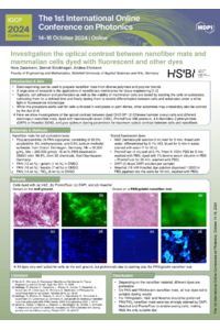
Electrospinning can be used to prepare nanofiber mats from diverse polymers and polymer blends with embedded metallic, ceramic, or other types of nanoparticles. Such nanofiber mats have been investigated for diverse applications, such as filtration, batteries, and supercapacitors; food packaging; or biomedicine and biotechnology. A large area of research is the application of nanofibrous membranes in tissue engineering. Typically, the cell adhesion and proliferation as well as the viability of mammalian cells are tested by seeding the cells on the substrates under examination, cultivating them for a defined period of time, and finally dyeing them to enable differentiation between cells and substrates under a white light or fluorescence microscope. While this procedure works well for cells cultivated in well plates or petri dishes, other substrates may undesirably also be colored by the dye. Here, we show investigations of the optical contrast between dyed CHO DP-12 (Chinese hamster ovary) cells and different nanofiber mats electrospun from pure poly(acrylonitrile) (PAN) as well as PAN/gelatin, PAN/keratin, and PAN/TiO2. After cultivation for 5 days, the cells were fixed with glyoxal and afterwards dyed with haematoxylin–eosin (H&E), PromoFluor 488 premium, 4,6-diamidino-2-phenylindole (DAPI), or Hoechst 33342. Examination by white light or fluorescence microscopy, respectively, revealed that, in particular, the PAN/gelatin nanofiber mats—which were the most advantageous regarding cell growth—were also colored by the dyes. This paper reports the optimized dyeing parameters to maximize the optical contrast between CHO cells and nanofiber mats.
Keywords
mammalian cells
CHO cells
electrospinning
tissue engineering
fluorescent dyes
Poster
095838 - Poster.PDF
Prototype of a Public Computer System with Fast Automatic Touchscreen Disinfection by Integrated UVC LEDs and Total Reflection
Four-Frame Structured Illumination Microscopy Based on Checkerboard Pattern