Event submissions
Published
This submission belongs to the session 2. Biomechanics and Sports Medicine of the event The 1st International Online Conference on Bioengineering
Published date
15 Oct, 2024
Academic Editor
'%3e%3cpath%20d='M12.6657%2010.9104C12.4185%2010.325%2012.0599%209.79327%2011.6097%209.34482C11.1609%208.89508%2010.6293%208.5365%2010.0442%208.28888C10.0389%208.28626%2010.0337%208.28495%2010.0285%208.28233C10.8446%207.69279%2011.3752%206.73249%2011.3752%205.64904C11.3752%203.8542%209.92103%202.39999%208.1262%202.39999C6.33136%202.39999%204.87715%203.8542%204.87715%205.64904C4.87715%206.73249%205.40774%207.69279%206.22393%208.28364C6.21869%208.28626%206.21345%208.28757%206.20821%208.29019C5.62129%208.5378%205.09463%208.89284%204.64265%209.34613C4.1929%209.79493%203.83432%2010.3266%203.58671%2010.9117C3.34345%2011.4845%203.21226%2012.0987%203.20023%2012.7209C3.19988%2012.7349%203.20233%2012.7488%203.20744%2012.7619C3.21255%2012.7749%203.22022%2012.7867%203.22998%2012.7968C3.23975%2012.8068%203.25142%2012.8147%203.26431%2012.8202C3.2772%2012.8256%203.29105%2012.8284%203.30504%2012.8284H4.09109C4.14874%2012.8284%204.19459%2012.7825%204.1959%2012.7262C4.2221%2011.7148%204.62823%2010.7676%205.34617%2010.0497C6.08899%209.30683%207.0755%208.89808%208.1262%208.89808C9.17689%208.89808%2010.1634%209.30683%2010.9062%2010.0497C11.6242%2010.7676%2012.0303%2011.7148%2012.0565%2012.7262C12.0578%2012.7838%2012.1037%2012.8284%2012.1613%2012.8284H12.9474C12.9613%2012.8284%2012.9752%2012.8256%2012.9881%2012.8202C13.001%2012.8147%2013.0126%2012.8068%2013.0224%2012.7968C13.0322%2012.7867%2013.0398%2012.7749%2013.0449%2012.7619C13.0501%2012.7488%2013.0525%2012.7349%2013.0522%2012.7209C13.0391%2012.0947%2012.9094%2011.4855%2012.6657%2010.9104ZM8.1262%207.9024C7.52486%207.9024%206.9589%207.6679%206.53312%207.24211C6.10733%206.81633%205.87283%206.25037%205.87283%205.64904C5.87283%205.0477%206.10733%204.48174%206.53312%204.05596C6.9589%203.63018%207.52486%203.39567%208.1262%203.39567C8.72753%203.39567%209.29349%203.63018%209.71927%204.05596C10.1451%204.48174%2010.3796%205.0477%2010.3796%205.64904C10.3796%206.25037%2010.1451%206.81633%209.71927%207.24211C9.29349%207.6679%208.72753%207.9024%208.1262%207.9024Z'%20fill='%235D1EE1'/%3e%3c/g%3e%3c/svg%3e) Franz Konstantin Fuss
Franz Konstantin FussCitation
Rui Pedro Pereira de Almeida, Manuel Lobo, Sérgio Tavares, Carlos da Silva, Ana Bárbara, Nélia Gaudêncio, Magda Ramos, António Abrantes, Advancements in Medical Imaging Methods for Body Composition Assessment: A Literature Review, in Proceedings of The 1st International Online Conference on Bioengineering, 16 October–18 October 2024, MDPI: Basel, Switzerland
Share
Email
Facebook
Twitter
LinkedIn
Advancements in Medical Imaging Methods for Body Composition Assessment: A Literature Review
'%3e%3cpath%20d='M12.6647%2010.9104C12.4176%2010.325%2012.0589%209.7933%2011.6088%209.34485C11.16%208.89511%2010.6283%208.53653%2010.0432%208.28891C10.038%208.28629%2010.0327%208.28498%2010.0275%208.28236C10.8437%207.69282%2011.3743%206.73252%2011.3743%205.64907C11.3743%203.85423%209.92005%202.40002%208.12522%202.40002C6.33038%202.40002%204.87618%203.85423%204.87618%205.64907C4.87618%206.73252%205.40677%207.69282%206.22296%208.28367C6.21772%208.28629%206.21248%208.2876%206.20724%208.29022C5.62031%208.53783%205.09365%208.89287%204.64167%209.34616C4.19192%209.79496%203.83334%2010.3266%203.58573%2010.9117C3.34247%2011.4846%203.21128%2012.0987%203.19925%2012.721C3.1989%2012.735%203.20135%2012.7489%203.20646%2012.7619C3.21157%2012.7749%203.21924%2012.7868%203.22901%2012.7968C3.23877%2012.8068%203.25045%2012.8148%203.26334%2012.8202C3.27623%2012.8256%203.29007%2012.8284%203.30406%2012.8284H4.09012C4.14776%2012.8284%204.19362%2012.7825%204.19493%2012.7262C4.22113%2011.7148%204.62726%2010.7676%205.34519%2010.0497C6.08802%209.30686%207.07452%208.89811%208.12522%208.89811C9.17592%208.89811%2010.1624%209.30686%2010.9052%2010.0497C11.6232%2010.7676%2012.0293%2011.7148%2012.0555%2012.7262C12.0568%2012.7839%2012.1027%2012.8284%2012.1603%2012.8284H12.9464C12.9604%2012.8284%2012.9742%2012.8256%2012.9871%2012.8202C13%2012.8148%2013.0117%2012.8068%2013.0214%2012.7968C13.0312%2012.7868%2013.0389%2012.7749%2013.044%2012.7619C13.0491%2012.7489%2013.0515%2012.735%2013.0512%2012.721C13.0381%2012.0947%2012.9084%2011.4856%2012.6647%2010.9104ZM8.12522%207.90243C7.52388%207.90243%206.95792%207.66793%206.53214%207.24215C6.10636%206.81636%205.87185%206.2504%205.87185%205.64907C5.87185%205.04773%206.10636%204.48177%206.53214%204.05599C6.95792%203.63021%207.52388%203.3957%208.12522%203.3957C8.72655%203.3957%209.29252%203.63021%209.7183%204.05599C10.1441%204.48177%2010.3786%205.04773%2010.3786%205.64907C10.3786%206.2504%2010.1441%206.81636%209.7183%207.24215C9.29252%207.66793%208.72655%207.90243%208.12522%207.90243Z'%20fill='%235D1EE1'/%3e%3c/g%3e%3c/svg%3e)
Rui Pedro Pereira de Almeida 1,2
Manuel Lobo 3,4
Sérgio Tavares 5,6

Carlos da Silva 7
Ana Bárbara 6
Nélia Gaudêncio 6
Magda Ramos 6
António Abrantes 2,6,8
1. University of Algarve, Campus de Gambelas, 8005-139, Faro, Portugal
2. Comprehensive Health Research Center, University of Evora, Portugal
3. Local Health Unit of the Northeast, Portugal
4. Polytechnic University of Castelo Branco, Portugal
5. Algarve Local Health Unit, Basic Emergency Center of Vila Real de Santo António, Portugal
6. University of Algarve, Portugal
7. University of Évora, Portugal
8. CICS.NOVA.UÉvora, Portugal
Abstract
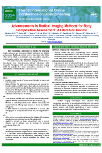
Introduction: Accurate body composition assessment is critical in clinical and sports settings to assess health, fitness, and disease risk. Advances in medical imaging technology have considerably increased the ability to measure and analyze body composition. This review examines these imaging techniques, focusing on their application in sports medicine. Methods: This review covers current imaging techniques for body composition assessment, including Dual-Energy X-ray Absorptiometry (DXA), Magnetic Resonance Imaging (MRI), Computed Tomography (CT), and Ultrasound (US). Parameters such as accuracy, precision, application range, and radiation exposure were evaluated. Data consistency was ensured by cross-referencing findings from multiple sources and by prioritizing analyses with large sample sizes and rigorous methods. Results and Discussion: DXA is widely recognized for its high accuracy and low radiation efficiency, providing comprehensive data on bone, lean mass, and fat mass. It is specifically useful for detailed regional analysis, although hydration status can affect its accuracy. MRI provides detailed information without ionizing radiation, and is ideal for monitoring muscle health and detecting sarcopenia, despite its high cost and limited availability. CT provides detailed cross-sectional images for precise tissue measurement but involves higher radiation exposure. Ultrasound is a practical, non-invasive, cost-effective method for assessing subcutaneous fat and muscle thickness, though less detailed and more operator-dependent compared to MRI and CT. Conclusions: Medical imaging technologies have greatly improved body composition assessment, providing detailed insights into muscle and fat distribution. DXA stands out as the gold standard for its balance of accuracy, safety, and cost balance. MRI and CT provide detailed imaging but come with high costs and radiation exposure. Ultrasound remains a practical alternative for early evaluation, though less detailed, assessments. Continuous technological developments and artificial intelligence promise to further advance these approaches in sports medicine, leading to better health and performance for athletes.
Keywords
Body Composition Assessment
Medical Imaging
Dual-Energy X-ray Absorptiometry
Magnetic Resonance Imaging
Ultrasound
Sports Medicine
Poster
IOCBE_Poster_sciforum-101083_V1FINAL.pdf
Review on the formulation of cellulose-based hydrogels and their biomedical applications
NeuroAmph: Innovative Synergy of Polydopamine and Peptide Amphiphiles for Enhanced Cognitive Pathology Treatment