Event submissions
Published

This submission belongs to the session S2. Physical Sensors of the event The 11th International Electronic Conference on Sensors and Applications
Published date
26 Nov, 2024
Academic Editor
'%3e%3cpath%20d='M12.6657%2010.9104C12.4185%2010.325%2012.0599%209.79327%2011.6097%209.34482C11.1609%208.89508%2010.6293%208.5365%2010.0442%208.28888C10.0389%208.28626%2010.0337%208.28495%2010.0285%208.28233C10.8446%207.69279%2011.3752%206.73249%2011.3752%205.64904C11.3752%203.8542%209.92103%202.39999%208.1262%202.39999C6.33136%202.39999%204.87715%203.8542%204.87715%205.64904C4.87715%206.73249%205.40774%207.69279%206.22393%208.28364C6.21869%208.28626%206.21345%208.28757%206.20821%208.29019C5.62129%208.5378%205.09463%208.89284%204.64265%209.34613C4.1929%209.79493%203.83432%2010.3266%203.58671%2010.9117C3.34345%2011.4845%203.21226%2012.0987%203.20023%2012.7209C3.19988%2012.7349%203.20233%2012.7488%203.20744%2012.7619C3.21255%2012.7749%203.22022%2012.7867%203.22998%2012.7968C3.23975%2012.8068%203.25142%2012.8147%203.26431%2012.8202C3.2772%2012.8256%203.29105%2012.8284%203.30504%2012.8284H4.09109C4.14874%2012.8284%204.19459%2012.7825%204.1959%2012.7262C4.2221%2011.7148%204.62823%2010.7676%205.34617%2010.0497C6.08899%209.30683%207.0755%208.89808%208.1262%208.89808C9.17689%208.89808%2010.1634%209.30683%2010.9062%2010.0497C11.6242%2010.7676%2012.0303%2011.7148%2012.0565%2012.7262C12.0578%2012.7838%2012.1037%2012.8284%2012.1613%2012.8284H12.9474C12.9613%2012.8284%2012.9752%2012.8256%2012.9881%2012.8202C13.001%2012.8147%2013.0126%2012.8068%2013.0224%2012.7968C13.0322%2012.7867%2013.0398%2012.7749%2013.0449%2012.7619C13.0501%2012.7488%2013.0525%2012.7349%2013.0522%2012.7209C13.0391%2012.0947%2012.9094%2011.4855%2012.6657%2010.9104ZM8.1262%207.9024C7.52486%207.9024%206.9589%207.6679%206.53312%207.24211C6.10733%206.81633%205.87283%206.25037%205.87283%205.64904C5.87283%205.0477%206.10733%204.48174%206.53312%204.05596C6.9589%203.63018%207.52486%203.39567%208.1262%203.39567C8.72753%203.39567%209.29349%203.63018%209.71927%204.05596C10.1451%204.48174%2010.3796%205.0477%2010.3796%205.64904C10.3796%206.25037%2010.1451%206.81633%209.71927%207.24211C9.29349%207.6679%208.72753%207.9024%208.1262%207.9024Z'%20fill='%235D1EE1'/%3e%3c/g%3e%3c/svg%3e) Francisco Falcone
Francisco FalconeCitation
Bahar Faramarzi, Marianna Portaccio, Lorenzo Manti, Maria Daniela Falco, Manuela Iezzi, Maria Lepore, Discrimination of different human cell lines by using FT-IR spectra spectroscopy, in Proceedings of The 11th International Electronic Conference on Sensors and Applications, 26 November–28 November 2024, MDPI: Basel, Switzerland, doi: 10.3390/ecsa-11-20499
Share
Email
Facebook
Twitter
LinkedIn
Discrimination of different human cell lines by using FT-IR spectra spectroscopy
'%3e%3cpath%20d='M12.6647%2010.9104C12.4176%2010.325%2012.0589%209.7933%2011.6088%209.34485C11.16%208.89511%2010.6283%208.53653%2010.0432%208.28891C10.038%208.28629%2010.0327%208.28498%2010.0275%208.28236C10.8437%207.69282%2011.3743%206.73252%2011.3743%205.64907C11.3743%203.85423%209.92005%202.40002%208.12522%202.40002C6.33038%202.40002%204.87618%203.85423%204.87618%205.64907C4.87618%206.73252%205.40677%207.69282%206.22296%208.28367C6.21772%208.28629%206.21248%208.2876%206.20724%208.29022C5.62031%208.53783%205.09365%208.89287%204.64167%209.34616C4.19192%209.79496%203.83334%2010.3266%203.58573%2010.9117C3.34247%2011.4846%203.21128%2012.0987%203.19925%2012.721C3.1989%2012.735%203.20135%2012.7489%203.20646%2012.7619C3.21157%2012.7749%203.21924%2012.7868%203.22901%2012.7968C3.23877%2012.8068%203.25045%2012.8148%203.26334%2012.8202C3.27623%2012.8256%203.29007%2012.8284%203.30406%2012.8284H4.09012C4.14776%2012.8284%204.19362%2012.7825%204.19493%2012.7262C4.22113%2011.7148%204.62726%2010.7676%205.34519%2010.0497C6.08802%209.30686%207.07452%208.89811%208.12522%208.89811C9.17592%208.89811%2010.1624%209.30686%2010.9052%2010.0497C11.6232%2010.7676%2012.0293%2011.7148%2012.0555%2012.7262C12.0568%2012.7839%2012.1027%2012.8284%2012.1603%2012.8284H12.9464C12.9604%2012.8284%2012.9742%2012.8256%2012.9871%2012.8202C13%2012.8148%2013.0117%2012.8068%2013.0214%2012.7968C13.0312%2012.7868%2013.0389%2012.7749%2013.044%2012.7619C13.0491%2012.7489%2013.0515%2012.735%2013.0512%2012.721C13.0381%2012.0947%2012.9084%2011.4856%2012.6647%2010.9104ZM8.12522%207.90243C7.52388%207.90243%206.95792%207.66793%206.53214%207.24215C6.10636%206.81636%205.87185%206.2504%205.87185%205.64907C5.87185%205.04773%206.10636%204.48177%206.53214%204.05599C6.95792%203.63021%207.52388%203.3957%208.12522%203.3957C8.72655%203.3957%209.29252%203.63021%209.7183%204.05599C10.1441%204.48177%2010.3786%205.04773%2010.3786%205.64907C10.3786%206.2504%2010.1441%206.81636%209.7183%207.24215C9.29252%207.66793%208.72655%207.90243%208.12522%207.90243Z'%20fill='%235D1EE1'/%3e%3c/g%3e%3c/svg%3e)
Bahar Faramarzi 1
Marianna Portaccio 1

Lorenzo Manti 2,3
Maria Daniela Falco 4
Manuela Iezzi 5,6

Maria Lepore 1
1. Dipartimento di Medicina Sperimentale, Università della Campania “Luigi Vanvitelli”, 80138 Napoli, Italy;
2. Dipartimento di Fisica “E. Pancini”, Università degli Studi di Napoli “Federico II”, 80126 Napoli, Italy;
3. Istituto Nazionale di Fisica Nucleare—Sezione di Napoli, 80100 Napoli, Italy;
4. ASL2 Lanciano-Vasto-Chieti, UOSD di Fisica Sanitaria, 66100 Chieti, Italy;
5. Department of Neurosciences, Imaging and Clinical Sciences, Center for Advanced Studies and Technnology (CAST) “G. d’Annunzio” University of Chieti-Pescara, 66100 Chieti, Italy;
6. ASL2 Lanciano-Vasto-Chieti, UOC Anatomia e Istologia Patologica, 66100 Chieti, Italy.
Abstract
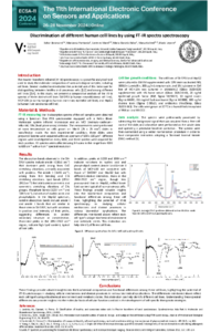
Fourier Transform Infrared (FT-IR) spectroscopy is a powerful analytical technique to obtain molecular fingerprints of various biological samples. Several studies have indicated the potential use of this technique for distinguishing between healthy and cancerous cells [1,2] and among different cell lines [3,4]. This study aims to compare the FT-IR spectra of three distinct cell lines, SH-SY5Y (neuroblastoma), HepG2 (hepatocellular carcinoma), and MCF-10A (epithelial mammary), to identify characteristic features in their spectra that can be used for this purpose. The cells were grown on MirrIR substrates, and the spectra were acquired in transflection mode using the microscopic stage of a Perkin Elmer Spectrum One Spectrometer equipped with a mercury cadmium telluride (MCT) detector. The FTIR spectra revealed significant protein, lipid, and nucleic acid content variations among the cell lines. The differences mentioned above reflect each cell type's unique biochemical environment and metabolic states. This distinction can help identify different cell lines. Understanding these spectral differences can provide insights into the molecular basis of cellular functions and aid in the development of cell-specific therapeutic strategies.
References
[1] M. Lasalvia, V. Capozzi, G. Classification of healthy and cancerous colon cells by Fourier transform infrared spectroscopy Spectrochimica Acta Part A: Molecular and Biomolecular Spectroscopy 321 (2024) 124683
[2] W.M. Elshemey, A.M. Ismail, N.S. Elbialy, Molecular-level characterization of normal, benign, and malignant breast tissues using FTIR spectroscopy, J. Med. Biol. Eng. 36 (2016) 369–378.
[3] Zendehdel R, H Shirazi F. Discrimination of Human Cell Lines by Infrared Spectroscopy and Mathematical Modeling. Iran J Pharm Res. 2015 Summer;14(3):803-10.
[4] Yadi Wang, Wentao Dai, Zhixiao Liu, Jixiang Liu, Jie Cheng, Yuanyuan Li, Xueling Li, Jun Hu, and Junhong Lü Single-Cell Infrared Microspectroscopy Quantifies Dynamic Heterogeneity of Mesenchymal Stem Cells during Adipogenic Differentiation Analytical Chemistry 2021 93 (2), 671-676.
Keywords
Human cell lines
SH-SY5Y
HepG2
MCF-10A
FT-IR spectroscopy
Proteins
Lipids
DNA
Cell identification.
Manuscript
engproc-sciforum_101277_S2.pdf
Poster
Poster-101277 (ECSA-11).pdf
Robosim: Design, Implementation, and Applications of a Line Follower Robot Simulator
A Wearable Reflectance PPG Optical Sensor Enabling Contact Pressure and Skin Temperature Measurement