Event submissions
Published
This submission belongs to the session S6. Plant–Crop Biology and Biochemistry of the event The 4th International Electronic Conference on Agronomy
Published date
29 Nov, 2024
Academic Editor
'%3e%3cpath%20d='M12.6657%2010.9104C12.4185%2010.325%2012.0599%209.79327%2011.6097%209.34482C11.1609%208.89508%2010.6293%208.5365%2010.0442%208.28888C10.0389%208.28626%2010.0337%208.28495%2010.0285%208.28233C10.8446%207.69279%2011.3752%206.73249%2011.3752%205.64904C11.3752%203.8542%209.92103%202.39999%208.1262%202.39999C6.33136%202.39999%204.87715%203.8542%204.87715%205.64904C4.87715%206.73249%205.40774%207.69279%206.22393%208.28364C6.21869%208.28626%206.21345%208.28757%206.20821%208.29019C5.62129%208.5378%205.09463%208.89284%204.64265%209.34613C4.1929%209.79493%203.83432%2010.3266%203.58671%2010.9117C3.34345%2011.4845%203.21226%2012.0987%203.20023%2012.7209C3.19988%2012.7349%203.20233%2012.7488%203.20744%2012.7619C3.21255%2012.7749%203.22022%2012.7867%203.22998%2012.7968C3.23975%2012.8068%203.25142%2012.8147%203.26431%2012.8202C3.2772%2012.8256%203.29105%2012.8284%203.30504%2012.8284H4.09109C4.14874%2012.8284%204.19459%2012.7825%204.1959%2012.7262C4.2221%2011.7148%204.62823%2010.7676%205.34617%2010.0497C6.08899%209.30683%207.0755%208.89808%208.1262%208.89808C9.17689%208.89808%2010.1634%209.30683%2010.9062%2010.0497C11.6242%2010.7676%2012.0303%2011.7148%2012.0565%2012.7262C12.0578%2012.7838%2012.1037%2012.8284%2012.1613%2012.8284H12.9474C12.9613%2012.8284%2012.9752%2012.8256%2012.9881%2012.8202C13.001%2012.8147%2013.0126%2012.8068%2013.0224%2012.7968C13.0322%2012.7867%2013.0398%2012.7749%2013.0449%2012.7619C13.0501%2012.7488%2013.0525%2012.7349%2013.0522%2012.7209C13.0391%2012.0947%2012.9094%2011.4855%2012.6657%2010.9104ZM8.1262%207.9024C7.52486%207.9024%206.9589%207.6679%206.53312%207.24211C6.10733%206.81633%205.87283%206.25037%205.87283%205.64904C5.87283%205.0477%206.10733%204.48174%206.53312%204.05596C6.9589%203.63018%207.52486%203.39567%208.1262%203.39567C8.72753%203.39567%209.29349%203.63018%209.71927%204.05596C10.1451%204.48174%2010.3796%205.0477%2010.3796%205.64904C10.3796%206.25037%2010.1451%206.81633%209.71927%207.24211C9.29349%207.6679%208.72753%207.9024%208.1262%207.9024Z'%20fill='%235D1EE1'/%3e%3c/g%3e%3c/svg%3e) Monica Boscaiu
Monica BoscaiuCitation
Binsy Karattuchali, Sharon Aravind, Sivaranjani R, Farsana Soudath K P, Comparative anatomical and biochemical studies of in vitro and in vivo plants of ginger, in Proceedings of The 4th International Electronic Conference on Agronomy, 2 December–5 December 2024, MDPI: Basel, Switzerland
Share
Email
Facebook
Twitter
LinkedIn
Comparative anatomical and biochemical studies of in vitro and in vivo plants of ginger
'%3e%3cpath%20d='M12.6647%2010.9104C12.4176%2010.325%2012.0589%209.7933%2011.6088%209.34485C11.16%208.89511%2010.6283%208.53653%2010.0432%208.28891C10.038%208.28629%2010.0327%208.28498%2010.0275%208.28236C10.8437%207.69282%2011.3743%206.73252%2011.3743%205.64907C11.3743%203.85423%209.92005%202.40002%208.12522%202.40002C6.33038%202.40002%204.87618%203.85423%204.87618%205.64907C4.87618%206.73252%205.40677%207.69282%206.22296%208.28367C6.21772%208.28629%206.21248%208.2876%206.20724%208.29022C5.62031%208.53783%205.09365%208.89287%204.64167%209.34616C4.19192%209.79496%203.83334%2010.3266%203.58573%2010.9117C3.34247%2011.4846%203.21128%2012.0987%203.19925%2012.721C3.1989%2012.735%203.20135%2012.7489%203.20646%2012.7619C3.21157%2012.7749%203.21924%2012.7868%203.22901%2012.7968C3.23877%2012.8068%203.25045%2012.8148%203.26334%2012.8202C3.27623%2012.8256%203.29007%2012.8284%203.30406%2012.8284H4.09012C4.14776%2012.8284%204.19362%2012.7825%204.19493%2012.7262C4.22113%2011.7148%204.62726%2010.7676%205.34519%2010.0497C6.08802%209.30686%207.07452%208.89811%208.12522%208.89811C9.17592%208.89811%2010.1624%209.30686%2010.9052%2010.0497C11.6232%2010.7676%2012.0293%2011.7148%2012.0555%2012.7262C12.0568%2012.7839%2012.1027%2012.8284%2012.1603%2012.8284H12.9464C12.9604%2012.8284%2012.9742%2012.8256%2012.9871%2012.8202C13%2012.8148%2013.0117%2012.8068%2013.0214%2012.7968C13.0312%2012.7868%2013.0389%2012.7749%2013.044%2012.7619C13.0491%2012.7489%2013.0515%2012.735%2013.0512%2012.721C13.0381%2012.0947%2012.9084%2011.4856%2012.6647%2010.9104ZM8.12522%207.90243C7.52388%207.90243%206.95792%207.66793%206.53214%207.24215C6.10636%206.81636%205.87185%206.2504%205.87185%205.64907C5.87185%205.04773%206.10636%204.48177%206.53214%204.05599C6.95792%203.63021%207.52388%203.3957%208.12522%203.3957C8.72655%203.3957%209.29252%203.63021%209.7183%204.05599C10.1441%204.48177%2010.3786%205.04773%2010.3786%205.64907C10.3786%206.2504%2010.1441%206.81636%209.7183%207.24215C9.29252%207.66793%208.72655%207.90243%208.12522%207.90243Z'%20fill='%235D1EE1'/%3e%3c/g%3e%3c/svg%3e)
Binsy Karattuchali 1
Sharon Aravind 1
Sivaranjani R 2
Farsana Soudath K P 1
1. Division of Crop Improvement and Biotechnology, ICAR - Indian Institute of Spices Research, Kozhikode 673012, Kerala, India
2. Division of Crop Production and Post Harvest Technology, ICAR - Indian Institute of Spices Research, Kozhikode 673012, Kerala, India
Abstract
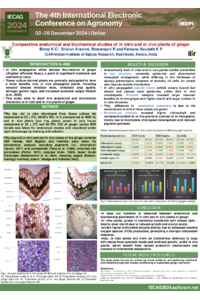
In vitro propagation offers disease-free material in ginger (Zingiber officinale Rosc.), a plant of significant medicinal and commercial value. This study aims to investigate the anatomical and biochemical characteristics of in vitro and in vivo plants of ginger. Fifty-day-old in vitro (developed from tissue culture lab maintained at 25 ± 2°C and 90-92% RH over 14 h photoperiod at 3000 lx) and in vivo plants (pro-tray plants grown in polyhouse maintained at 25 ± 2°C and 60-70% RH) of the ginger variety IISR Varada were taken to be studied at the ICAR—Indian Institute of Spices Research, Kerala, India. Transverse sections of leaves, the pseudo-stem, and the rhizomes of the in vitro and in vivo ginger plants were taken and visualized under light microscopy.
In vitro and in vivo ginger possess similar leaf structures but differ in their spongy parenchyma thickness, the number of stomata, oil cells, and air canals, and their vascular bundle distribution. The in vitro pseudo-stems had a closely bound leaf sheath and epidermis unlike their in vivo counterparts. Rhizome analysis revealed larger vascular bundles in the in vivo ginger (normal rhizomes produced at farm) and higher starch and sugar content in the in vitro rhizomes (micro-rhizomes developed at tissue culture lab).
Biochemical analysis revealed that the total chlorophyll and carotenoid content was significantly higher in the in vivo ginger (1.1989 mg/g and 67.24 mg/g) compared to the in vitro ginger (0.7173 mg/g and 50.87 mg/g, respectively). Furthermore, the enzyme activities of peroxidase (0.063 mg/g), catalase (108.248 U/g), and superoxide dismutase (0.367 U/g); the starch content (22.84%); and the total soluble sugars (0.093%) were higher in the in vitro ginger plants.
Hence, the anatomical and biochemical variations observed in the different parts of the in vitro and in vivo ginger plants may be due to the differences in the growing conditions and the media used in the in vitro conditions.
Keywords
in vitro
in vivo
epidermis
vascular bundle
starch grains
pseudostem
rhizome
translocation
Chlorophyll content
Carotenoid levels
Enzyme activities
Starch content
Total soluble sugars
Poster
Poster-.pdf
An in silico approach for the comparative characterization of imidazolonepropionase from Agrobacterium fabrum and Bacillus subtilis: an imperative enzyme for histidine degradation
Germinability and electrical conductivity of seeds of groundnut varieties