Event submissions
Published
This submission belongs to the session S3. Computing and Artificial Intelligence of the event The 5th International Electronic Conference on Applied Sciences
Published date
04 Dec, 2024
Academic Editor
'%3e%3cpath%20d='M12.6657%2010.9104C12.4185%2010.325%2012.0599%209.79327%2011.6097%209.34482C11.1609%208.89508%2010.6293%208.5365%2010.0442%208.28888C10.0389%208.28626%2010.0337%208.28495%2010.0285%208.28233C10.8446%207.69279%2011.3752%206.73249%2011.3752%205.64904C11.3752%203.8542%209.92103%202.39999%208.1262%202.39999C6.33136%202.39999%204.87715%203.8542%204.87715%205.64904C4.87715%206.73249%205.40774%207.69279%206.22393%208.28364C6.21869%208.28626%206.21345%208.28757%206.20821%208.29019C5.62129%208.5378%205.09463%208.89284%204.64265%209.34613C4.1929%209.79493%203.83432%2010.3266%203.58671%2010.9117C3.34345%2011.4845%203.21226%2012.0987%203.20023%2012.7209C3.19988%2012.7349%203.20233%2012.7488%203.20744%2012.7619C3.21255%2012.7749%203.22022%2012.7867%203.22998%2012.7968C3.23975%2012.8068%203.25142%2012.8147%203.26431%2012.8202C3.2772%2012.8256%203.29105%2012.8284%203.30504%2012.8284H4.09109C4.14874%2012.8284%204.19459%2012.7825%204.1959%2012.7262C4.2221%2011.7148%204.62823%2010.7676%205.34617%2010.0497C6.08899%209.30683%207.0755%208.89808%208.1262%208.89808C9.17689%208.89808%2010.1634%209.30683%2010.9062%2010.0497C11.6242%2010.7676%2012.0303%2011.7148%2012.0565%2012.7262C12.0578%2012.7838%2012.1037%2012.8284%2012.1613%2012.8284H12.9474C12.9613%2012.8284%2012.9752%2012.8256%2012.9881%2012.8202C13.001%2012.8147%2013.0126%2012.8068%2013.0224%2012.7968C13.0322%2012.7867%2013.0398%2012.7749%2013.0449%2012.7619C13.0501%2012.7488%2013.0525%2012.7349%2013.0522%2012.7209C13.0391%2012.0947%2012.9094%2011.4855%2012.6657%2010.9104ZM8.1262%207.9024C7.52486%207.9024%206.9589%207.6679%206.53312%207.24211C6.10733%206.81633%205.87283%206.25037%205.87283%205.64904C5.87283%205.0477%206.10733%204.48174%206.53312%204.05596C6.9589%203.63018%207.52486%203.39567%208.1262%203.39567C8.72753%203.39567%209.29349%203.63018%209.71927%204.05596C10.1451%204.48174%2010.3796%205.0477%2010.3796%205.64904C10.3796%206.25037%2010.1451%206.81633%209.71927%207.24211C9.29349%207.6679%208.72753%207.9024%208.1262%207.9024Z'%20fill='%235D1EE1'/%3e%3c/g%3e%3c/svg%3e) Eugenio Vocaturo
Eugenio VocaturoCitation
Tiar Mohamed Lakhdar, Terki Nadjiba, Accurate Classification of Acute Lymphoblastic Leukemia Subtypes Using Stacked Ensemble Learning on Peripheral Blood Smear Images, in Proceedings of The 5th International Electronic Conference on Applied Sciences, 4 December–6 December 2024, MDPI: Basel, Switzerland
Share
Email
Facebook
Twitter
LinkedIn
Accurate Classification of Acute Lymphoblastic Leukemia Subtypes Using Stacked Ensemble Learning on Peripheral Blood Smear Images
'%3e%3cpath%20d='M12.6647%2010.9104C12.4176%2010.325%2012.0589%209.7933%2011.6088%209.34485C11.16%208.89511%2010.6283%208.53653%2010.0432%208.28891C10.038%208.28629%2010.0327%208.28498%2010.0275%208.28236C10.8437%207.69282%2011.3743%206.73252%2011.3743%205.64907C11.3743%203.85423%209.92005%202.40002%208.12522%202.40002C6.33038%202.40002%204.87618%203.85423%204.87618%205.64907C4.87618%206.73252%205.40677%207.69282%206.22296%208.28367C6.21772%208.28629%206.21248%208.2876%206.20724%208.29022C5.62031%208.53783%205.09365%208.89287%204.64167%209.34616C4.19192%209.79496%203.83334%2010.3266%203.58573%2010.9117C3.34247%2011.4846%203.21128%2012.0987%203.19925%2012.721C3.1989%2012.735%203.20135%2012.7489%203.20646%2012.7619C3.21157%2012.7749%203.21924%2012.7868%203.22901%2012.7968C3.23877%2012.8068%203.25045%2012.8148%203.26334%2012.8202C3.27623%2012.8256%203.29007%2012.8284%203.30406%2012.8284H4.09012C4.14776%2012.8284%204.19362%2012.7825%204.19493%2012.7262C4.22113%2011.7148%204.62726%2010.7676%205.34519%2010.0497C6.08802%209.30686%207.07452%208.89811%208.12522%208.89811C9.17592%208.89811%2010.1624%209.30686%2010.9052%2010.0497C11.6232%2010.7676%2012.0293%2011.7148%2012.0555%2012.7262C12.0568%2012.7839%2012.1027%2012.8284%2012.1603%2012.8284H12.9464C12.9604%2012.8284%2012.9742%2012.8256%2012.9871%2012.8202C13%2012.8148%2013.0117%2012.8068%2013.0214%2012.7968C13.0312%2012.7868%2013.0389%2012.7749%2013.044%2012.7619C13.0491%2012.7489%2013.0515%2012.735%2013.0512%2012.721C13.0381%2012.0947%2012.9084%2011.4856%2012.6647%2010.9104ZM8.12522%207.90243C7.52388%207.90243%206.95792%207.66793%206.53214%207.24215C6.10636%206.81636%205.87185%206.2504%205.87185%205.64907C5.87185%205.04773%206.10636%204.48177%206.53214%204.05599C6.95792%203.63021%207.52388%203.3957%208.12522%203.3957C8.72655%203.3957%209.29252%203.63021%209.7183%204.05599C10.1441%204.48177%2010.3786%205.04773%2010.3786%205.64907C10.3786%206.2504%2010.1441%206.81636%209.7183%207.24215C9.29252%207.66793%208.72655%207.90243%208.12522%207.90243Z'%20fill='%235D1EE1'/%3e%3c/g%3e%3c/svg%3e)
Tiar Mohamed Lakhdar 1
Terki Nadjiba 1
1. University Mohamed Khider Biskra, BP 145 RP، 07000, Algeria
Abstract
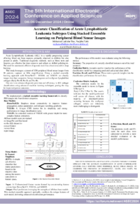
In this study, we leveraged a publicly available dataset containing 3256 peripheral blood smear (PBS) images, prepared in the bone marrow laboratory of Taleqani Hospital in Tehran, Iran. This dataset consists of blood samples from 89 patients suspected of Acute Lymphoblastic Leukemia (ALL). The images were captured using a Zeiss camera at 100x magnification and stored as JPG files. The dataset is divided into two primary classes: benign hematogones and malignant lymphoblasts. The malignant lymphoblasts are further categorized into three subtypes: Early Pre-B, Pre-B, and Pro-B ALL. The definitive classification of these cell types and subtypes was performed by a specialist using flow cytometry tools.
To classify these images into four distinct categories, we employed a stacked ensemble learning approach. Our model stack included three base models, DenseNet121, VGG16, and VGG19, with a K-Nearest Neighbors (KNN) classifier acting as the meta-model. This ensemble method capitalizes on the strengths of each individual model to improve overall classification performance. Our approach achieved a high accuracy of 94%, demonstrating its robustness and reliability in distinguishing between the various cell types and subtypes within the dataset.
The significant accuracy attained underscores the potential of advanced machine learning techniques in medical image analysis, particularly in the context of hematological malignancies. Our findings suggest that such methodologies could greatly enhance diagnostic precision and efficiency, leading to better patient outcomes. This study illustrates the promising application of deep learning models in the automated classification of ALL subtypes, paving the way for future advancements in the field.
Keywords
Machine learning
Deep learning
Cancer
Poster
Accurate Classification of Acute Lymphoblastic.pdf
Missing data imputation using machine learning techniques applied to IoT air quality sensors: A case study in Amazonia
Control of the spread of infectious diseases in cows on farms