Event submissions
Published
This submission belongs to the session C. Sustainable animal physiology and reproduction of the event The 3rd International Electronic Conference on Animals
Published date
07 Mar, 2025
Academic Editor
'%3e%3cpath%20d='M12.6657%2010.9104C12.4185%2010.325%2012.0599%209.79327%2011.6097%209.34482C11.1609%208.89508%2010.6293%208.5365%2010.0442%208.28888C10.0389%208.28626%2010.0337%208.28495%2010.0285%208.28233C10.8446%207.69279%2011.3752%206.73249%2011.3752%205.64904C11.3752%203.8542%209.92103%202.39999%208.1262%202.39999C6.33136%202.39999%204.87715%203.8542%204.87715%205.64904C4.87715%206.73249%205.40774%207.69279%206.22393%208.28364C6.21869%208.28626%206.21345%208.28757%206.20821%208.29019C5.62129%208.5378%205.09463%208.89284%204.64265%209.34613C4.1929%209.79493%203.83432%2010.3266%203.58671%2010.9117C3.34345%2011.4845%203.21226%2012.0987%203.20023%2012.7209C3.19988%2012.7349%203.20233%2012.7488%203.20744%2012.7619C3.21255%2012.7749%203.22022%2012.7867%203.22998%2012.7968C3.23975%2012.8068%203.25142%2012.8147%203.26431%2012.8202C3.2772%2012.8256%203.29105%2012.8284%203.30504%2012.8284H4.09109C4.14874%2012.8284%204.19459%2012.7825%204.1959%2012.7262C4.2221%2011.7148%204.62823%2010.7676%205.34617%2010.0497C6.08899%209.30683%207.0755%208.89808%208.1262%208.89808C9.17689%208.89808%2010.1634%209.30683%2010.9062%2010.0497C11.6242%2010.7676%2012.0303%2011.7148%2012.0565%2012.7262C12.0578%2012.7838%2012.1037%2012.8284%2012.1613%2012.8284H12.9474C12.9613%2012.8284%2012.9752%2012.8256%2012.9881%2012.8202C13.001%2012.8147%2013.0126%2012.8068%2013.0224%2012.7968C13.0322%2012.7867%2013.0398%2012.7749%2013.0449%2012.7619C13.0501%2012.7488%2013.0525%2012.7349%2013.0522%2012.7209C13.0391%2012.0947%2012.9094%2011.4855%2012.6657%2010.9104ZM8.1262%207.9024C7.52486%207.9024%206.9589%207.6679%206.53312%207.24211C6.10733%206.81633%205.87283%206.25037%205.87283%205.64904C5.87283%205.0477%206.10733%204.48174%206.53312%204.05596C6.9589%203.63018%207.52486%203.39567%208.1262%203.39567C8.72753%203.39567%209.29349%203.63018%209.71927%204.05596C10.1451%204.48174%2010.3796%205.0477%2010.3796%205.64904C10.3796%206.25037%2010.1451%206.81633%209.71927%207.24211C9.29349%207.6679%208.72753%207.9024%208.1262%207.9024Z'%20fill='%235D1EE1'/%3e%3c/g%3e%3c/svg%3e) Clive Julian Phillips
Clive Julian PhillipsCitation
Náyra Rachel Nascimento Luz, Pedro Augusto Pinheiro Brito, Gabriel Santos Costa Bezerra, Joana Letícia Cottin de Albuquerque, Euziele Oliveira de Santana, Romário Parente dos Santos, Luana Grasiele Pereira Bezerra, Andréia Maria da Silva, Alexsandra Fernandes Pereira, Nilza Dutra Alves, Moacir Franco de Oliveira, Alexandre Rodrigues Silva, Short-term in vitro culture of canine ovarian tissue aftercryopreservation by different techniques, in Proceedings of The 3rd International Electronic Conference on Animals, 12 March–14 March 2025, MDPI: Basel, Switzerland
Share
Email
Facebook
Twitter
LinkedIn
Short-term in vitro culture of canine ovarian tissue after
cryopreservation by different techniques
cryopreservation by different techniques
'%3e%3cpath%20d='M12.6647%2010.9104C12.4176%2010.325%2012.0589%209.7933%2011.6088%209.34485C11.16%208.89511%2010.6283%208.53653%2010.0432%208.28891C10.038%208.28629%2010.0327%208.28498%2010.0275%208.28236C10.8437%207.69282%2011.3743%206.73252%2011.3743%205.64907C11.3743%203.85423%209.92005%202.40002%208.12522%202.40002C6.33038%202.40002%204.87618%203.85423%204.87618%205.64907C4.87618%206.73252%205.40677%207.69282%206.22296%208.28367C6.21772%208.28629%206.21248%208.2876%206.20724%208.29022C5.62031%208.53783%205.09365%208.89287%204.64167%209.34616C4.19192%209.79496%203.83334%2010.3266%203.58573%2010.9117C3.34247%2011.4846%203.21128%2012.0987%203.19925%2012.721C3.1989%2012.735%203.20135%2012.7489%203.20646%2012.7619C3.21157%2012.7749%203.21924%2012.7868%203.22901%2012.7968C3.23877%2012.8068%203.25045%2012.8148%203.26334%2012.8202C3.27623%2012.8256%203.29007%2012.8284%203.30406%2012.8284H4.09012C4.14776%2012.8284%204.19362%2012.7825%204.19493%2012.7262C4.22113%2011.7148%204.62726%2010.7676%205.34519%2010.0497C6.08802%209.30686%207.07452%208.89811%208.12522%208.89811C9.17592%208.89811%2010.1624%209.30686%2010.9052%2010.0497C11.6232%2010.7676%2012.0293%2011.7148%2012.0555%2012.7262C12.0568%2012.7839%2012.1027%2012.8284%2012.1603%2012.8284H12.9464C12.9604%2012.8284%2012.9742%2012.8256%2012.9871%2012.8202C13%2012.8148%2013.0117%2012.8068%2013.0214%2012.7968C13.0312%2012.7868%2013.0389%2012.7749%2013.044%2012.7619C13.0491%2012.7489%2013.0515%2012.735%2013.0512%2012.721C13.0381%2012.0947%2012.9084%2011.4856%2012.6647%2010.9104ZM8.12522%207.90243C7.52388%207.90243%206.95792%207.66793%206.53214%207.24215C6.10636%206.81636%205.87185%206.2504%205.87185%205.64907C5.87185%205.04773%206.10636%204.48177%206.53214%204.05599C6.95792%203.63021%207.52388%203.3957%208.12522%203.3957C8.72655%203.3957%209.29252%203.63021%209.7183%204.05599C10.1441%204.48177%2010.3786%205.04773%2010.3786%205.64907C10.3786%206.2504%2010.1441%206.81636%209.7183%207.24215C9.29252%207.66793%208.72655%207.90243%208.12522%207.90243Z'%20fill='%235D1EE1'/%3e%3c/g%3e%3c/svg%3e)
Náyra Rachel Nascimento Luz 1
Pedro Augusto Pinheiro Brito 1
Gabriel Santos Costa Bezerra 1
Joana Letícia Cottin de Albuquerque 1
Euziele Oliveira de Santana 1
Romário Parente dos Santos 1
Luana Grasiele Pereira Bezerra 1
Andréia Maria da Silva 1
Alexsandra Fernandes Pereira 1
Nilza Dutra Alves 1
Moacir Franco de Oliveira 1

Alexandre Rodrigues Silva 1
1. Animal Germplasm Conservation Laboratory, Universidade Federal Rural do Semi-Árido, Mossoró - RN, 59.625-090, Brazil
Abstract
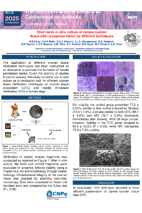
The application of different ovarian tissue vitrification techniques has been highlighted as an alternative to promote the formation of female germplasm banks. Given the scarcity of studies in canine species, this study aimed to use in vitro culture as an evaluation tool for different ovarian tissue vitrification techniques in female dogs. Five pairs of ovaries were collected from adult females. They were fragmented and subjected to vitrification using an ovarian tissue cryosystem (OTC) or adapted needle immersed vitrification (NIV). Fresh and vitrified fragments were cultured in vitro and evaluated for nine days for preantral follicle viability using a Trypan blue die and morphology through classic histology. The ultrastructural integrity of the ovarian tissue was checked by scanning electronic microscopy. The data were expressed as the mean and standard error and were compared using the Tukey test (P < 0.05). The fresh control group presented 77.2 ± 2.81% viability, similar to that of the control, cultured for 9 days (72.6 ± 1.8%), and also similar to both OTC (85.7 ± 3.6%) and NIV (75.7 ± 2.8%) treatments immediately after thawing. After 09 days of culture, however, viability in the OTC group dropped to 64.6 ± 16.2% (P< 0.05), while NIV maintained 72.8 ± 7.2% viability. For primordial preantral follicles morphology, there was no significant difference between the fresh control (85.5 ± 7.3%) and heated OTC (91.7 ± 5.4%), but after 09 days of culture, the OTC group (34.1 ± 9.0%) showed a significant decrease (P < 0.05), while NIV preserved their morphological integrity (63,6 ± 6,7 %). Regarding other follicle categories, no differences were observed among vitrification techniques. Through ultrastructural evaluation, disorganization patterns were observed for stromal cells of ovarian tissue at the use of OTC. In conclusion, the NIV technique allowed for a more efficient preservation of canine ovarian tissue than OTC.
Keywords
Biobank
Canids
Preantral follicles.
Poster
Banner Animals - Ovarian tissue - Pedro Brito.pdf
Comparative evaluation of sperm parameters of European and Africanized drones in Caatinga biome
Causal inference reveals long-term impact of health events on production performance in Holstein cattle