Event submissions
Published
This submission belongs to the session Session F. Cell Research in Animal Models of the event The 3rd International Online Conference on Cells
Published date
21 Mar, 2025
Academic Editor
'%3e%3cpath%20d='M12.6657%2010.9104C12.4185%2010.325%2012.0599%209.79327%2011.6097%209.34482C11.1609%208.89508%2010.6293%208.5365%2010.0442%208.28888C10.0389%208.28626%2010.0337%208.28495%2010.0285%208.28233C10.8446%207.69279%2011.3752%206.73249%2011.3752%205.64904C11.3752%203.8542%209.92103%202.39999%208.1262%202.39999C6.33136%202.39999%204.87715%203.8542%204.87715%205.64904C4.87715%206.73249%205.40774%207.69279%206.22393%208.28364C6.21869%208.28626%206.21345%208.28757%206.20821%208.29019C5.62129%208.5378%205.09463%208.89284%204.64265%209.34613C4.1929%209.79493%203.83432%2010.3266%203.58671%2010.9117C3.34345%2011.4845%203.21226%2012.0987%203.20023%2012.7209C3.19988%2012.7349%203.20233%2012.7488%203.20744%2012.7619C3.21255%2012.7749%203.22022%2012.7867%203.22998%2012.7968C3.23975%2012.8068%203.25142%2012.8147%203.26431%2012.8202C3.2772%2012.8256%203.29105%2012.8284%203.30504%2012.8284H4.09109C4.14874%2012.8284%204.19459%2012.7825%204.1959%2012.7262C4.2221%2011.7148%204.62823%2010.7676%205.34617%2010.0497C6.08899%209.30683%207.0755%208.89808%208.1262%208.89808C9.17689%208.89808%2010.1634%209.30683%2010.9062%2010.0497C11.6242%2010.7676%2012.0303%2011.7148%2012.0565%2012.7262C12.0578%2012.7838%2012.1037%2012.8284%2012.1613%2012.8284H12.9474C12.9613%2012.8284%2012.9752%2012.8256%2012.9881%2012.8202C13.001%2012.8147%2013.0126%2012.8068%2013.0224%2012.7968C13.0322%2012.7867%2013.0398%2012.7749%2013.0449%2012.7619C13.0501%2012.7488%2013.0525%2012.7349%2013.0522%2012.7209C13.0391%2012.0947%2012.9094%2011.4855%2012.6657%2010.9104ZM8.1262%207.9024C7.52486%207.9024%206.9589%207.6679%206.53312%207.24211C6.10733%206.81633%205.87283%206.25037%205.87283%205.64904C5.87283%205.0477%206.10733%204.48174%206.53312%204.05596C6.9589%203.63018%207.52486%203.39567%208.1262%203.39567C8.72753%203.39567%209.29349%203.63018%209.71927%204.05596C10.1451%204.48174%2010.3796%205.0477%2010.3796%205.64904C10.3796%206.25037%2010.1451%206.81633%209.71927%207.24211C9.29349%207.6679%208.72753%207.9024%208.1262%207.9024Z'%20fill='%235D1EE1'/%3e%3c/g%3e%3c/svg%3e) Alexander E. Kalyuzhny
Alexander E. KalyuzhnyCitation
Maria Eduarda Urzêda da Silva, Daniely Lisboa Matsnaka, Cristiane Damas Gil, Fernanda Regina Casagrande Giachini Vitorino, Sergio Marcelino de Oliveira, Kallyne Kioko Oliveira Mimura, Therapeutic properties of pequi lipophilic fraction (Caryocar brasiliense) in skin wound repair: modulation of inflammatory and proliferative phases, in Proceedings of The 3rd International Online Conference on Cells, 25 March–27 March 2025, MDPI: Basel, Switzerland
Share
Email
Facebook
Twitter
LinkedIn
Therapeutic properties of pequi lipophilic fraction (Caryocar brasiliense) in skin wound repair: modulation of inflammatory and proliferative phases
'%3e%3cpath%20d='M12.6647%2010.9104C12.4176%2010.325%2012.0589%209.7933%2011.6088%209.34485C11.16%208.89511%2010.6283%208.53653%2010.0432%208.28891C10.038%208.28629%2010.0327%208.28498%2010.0275%208.28236C10.8437%207.69282%2011.3743%206.73252%2011.3743%205.64907C11.3743%203.85423%209.92005%202.40002%208.12522%202.40002C6.33038%202.40002%204.87618%203.85423%204.87618%205.64907C4.87618%206.73252%205.40677%207.69282%206.22296%208.28367C6.21772%208.28629%206.21248%208.2876%206.20724%208.29022C5.62031%208.53783%205.09365%208.89287%204.64167%209.34616C4.19192%209.79496%203.83334%2010.3266%203.58573%2010.9117C3.34247%2011.4846%203.21128%2012.0987%203.19925%2012.721C3.1989%2012.735%203.20135%2012.7489%203.20646%2012.7619C3.21157%2012.7749%203.21924%2012.7868%203.22901%2012.7968C3.23877%2012.8068%203.25045%2012.8148%203.26334%2012.8202C3.27623%2012.8256%203.29007%2012.8284%203.30406%2012.8284H4.09012C4.14776%2012.8284%204.19362%2012.7825%204.19493%2012.7262C4.22113%2011.7148%204.62726%2010.7676%205.34519%2010.0497C6.08802%209.30686%207.07452%208.89811%208.12522%208.89811C9.17592%208.89811%2010.1624%209.30686%2010.9052%2010.0497C11.6232%2010.7676%2012.0293%2011.7148%2012.0555%2012.7262C12.0568%2012.7839%2012.1027%2012.8284%2012.1603%2012.8284H12.9464C12.9604%2012.8284%2012.9742%2012.8256%2012.9871%2012.8202C13%2012.8148%2013.0117%2012.8068%2013.0214%2012.7968C13.0312%2012.7868%2013.0389%2012.7749%2013.044%2012.7619C13.0491%2012.7489%2013.0515%2012.735%2013.0512%2012.721C13.0381%2012.0947%2012.9084%2011.4856%2012.6647%2010.9104ZM8.12522%207.90243C7.52388%207.90243%206.95792%207.66793%206.53214%207.24215C6.10636%206.81636%205.87185%206.2504%205.87185%205.64907C5.87185%205.04773%206.10636%204.48177%206.53214%204.05599C6.95792%203.63021%207.52388%203.3957%208.12522%203.3957C8.72655%203.3957%209.29252%203.63021%209.7183%204.05599C10.1441%204.48177%2010.3786%205.04773%2010.3786%205.64907C10.3786%206.2504%2010.1441%206.81636%209.7183%207.24215C9.29252%207.66793%208.72655%207.90243%208.12522%207.90243Z'%20fill='%235D1EE1'/%3e%3c/g%3e%3c/svg%3e)
Maria Eduarda Urzêda da Silva 1
Daniely Lisboa Matsnaka 1
Cristiane Damas Gil 2
Fernanda Regina Casagrande Giachini Vitorino 3
Sergio Marcelino de Oliveira 1,2
Kallyne Kioko Oliveira Mimura 2
1. Laboratory of Histophysiology and Animal Reproduction - Federal University of Mato Grosso - CUA/UFMT, Pontal do Araguaia 78698-000, MT, Brazil
2. Department of Morphology and Genetics, Paulista School of Medicine, Federal University of São Paulo (EPM-UNIFESP), São Paulo 04023-900, SP, Brazil
3. Laboratory of Vascular Biology and Histopathology - Federal University of Mato Grosso - CUA/UFMT, Barra do Garças 78605-091, MT, Brazil
Abstract
Introduction: Pequi (Caryocar brasiliense) is a fruit from Cerrado, Brazil, that has important therapeutic properties for the tissue repair of skin wounds. Methods: Balb/c mice were subjected to skin lesions and received daily topical treatment with the lipophilic fraction of pequi (Treated group) or saline solution (Control group). Animals from each group were sacrificed at 3, 7, and 14 days post injury (n=5). Macroscopic analyses were performed to assess wound contraction, and peripheral blood was collected for leukocyte quantification. Additionally, skin fragments were used for histopathological analysis (Hematoxylin–Eosin) and for quantification of the expression of the proteins Annexin A1 (AnxA1) and phosphorylated ERK (pERK) using the Western blot technique. CEUA-UFMT 23108.080623/2023-50. Results: At 3 days, the Treated animals exhibited a more robust scab on the lesion, providing greater physical and microbiological protection. At 7 days, wound contraction was significantly higher in the Treated group compared to the Control group (p ≤ 0.01), accelerating wound closure and preventing dehydration and infection risks. In peripheral blood, a higher number of neutrophils, eosinophils, and monocytes were identified in the Treated group compared to the Control group (p ≤ 0.0001; p ≤ 0.05; p ≤ 0.05, respectively), particularly during the early 3-day period. No difference was observed in AnxA1 protein expression between the experimental groups; however, the Treated group showed higher pERK expression at 7 days compared to the Control group (p ≤ 0.05). Conclusions: These data suggest that pequi oil acts as an effective modulator of the inflammatory phase, by inhibiting leukocyte transmigration, and of the proliferative phase, through the activation of the ERK pathway, preparing the tissue for regeneration without altering AnxA1 levels. Thus, it is evident that the lipophilic fraction of pequi accelerates tissue repair by modulating the inflammatory and proliferative phases, promoting more efficient healing. (Financial Support: Fapemat 000547-2023; CAPES 88887.976959/2024-00).
Keywords
Pequi
Skin
Healing
Inflammation.
Poster
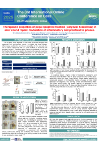
Banner Cells Conference 2025 - Silva et al..pdf
p75NTR modulation restores redox metabolism and blunts inflammation in a cell model of Rett syndrome
The effect of the lipophilic fraction of pequi on extracellular matrix remodeling in skin lesions in mice