Event submissions
Published
This submission belongs to the session S6. Antioxidants Extraction, Assay and Industrial Applications of the event The 2nd International Electronic Conference on Antioxidants
Published date
09 Apr, 2025
Academic Editor
'%3e%3cpath%20d='M12.6657%2010.9104C12.4185%2010.325%2012.0599%209.79327%2011.6097%209.34482C11.1609%208.89508%2010.6293%208.5365%2010.0442%208.28888C10.0389%208.28626%2010.0337%208.28495%2010.0285%208.28233C10.8446%207.69279%2011.3752%206.73249%2011.3752%205.64904C11.3752%203.8542%209.92103%202.39999%208.1262%202.39999C6.33136%202.39999%204.87715%203.8542%204.87715%205.64904C4.87715%206.73249%205.40774%207.69279%206.22393%208.28364C6.21869%208.28626%206.21345%208.28757%206.20821%208.29019C5.62129%208.5378%205.09463%208.89284%204.64265%209.34613C4.1929%209.79493%203.83432%2010.3266%203.58671%2010.9117C3.34345%2011.4845%203.21226%2012.0987%203.20023%2012.7209C3.19988%2012.7349%203.20233%2012.7488%203.20744%2012.7619C3.21255%2012.7749%203.22022%2012.7867%203.22998%2012.7968C3.23975%2012.8068%203.25142%2012.8147%203.26431%2012.8202C3.2772%2012.8256%203.29105%2012.8284%203.30504%2012.8284H4.09109C4.14874%2012.8284%204.19459%2012.7825%204.1959%2012.7262C4.2221%2011.7148%204.62823%2010.7676%205.34617%2010.0497C6.08899%209.30683%207.0755%208.89808%208.1262%208.89808C9.17689%208.89808%2010.1634%209.30683%2010.9062%2010.0497C11.6242%2010.7676%2012.0303%2011.7148%2012.0565%2012.7262C12.0578%2012.7838%2012.1037%2012.8284%2012.1613%2012.8284H12.9474C12.9613%2012.8284%2012.9752%2012.8256%2012.9881%2012.8202C13.001%2012.8147%2013.0126%2012.8068%2013.0224%2012.7968C13.0322%2012.7867%2013.0398%2012.7749%2013.0449%2012.7619C13.0501%2012.7488%2013.0525%2012.7349%2013.0522%2012.7209C13.0391%2012.0947%2012.9094%2011.4855%2012.6657%2010.9104ZM8.1262%207.9024C7.52486%207.9024%206.9589%207.6679%206.53312%207.24211C6.10733%206.81633%205.87283%206.25037%205.87283%205.64904C5.87283%205.0477%206.10733%204.48174%206.53312%204.05596C6.9589%203.63018%207.52486%203.39567%208.1262%203.39567C8.72753%203.39567%209.29349%203.63018%209.71927%204.05596C10.1451%204.48174%2010.3796%205.0477%2010.3796%205.64904C10.3796%206.25037%2010.1451%206.81633%209.71927%207.24211C9.29349%207.6679%208.72753%207.9024%208.1262%207.9024Z'%20fill='%235D1EE1'/%3e%3c/g%3e%3c/svg%3e) Alessandra Napolitano
Alessandra NapolitanoCitation
Ragavi Ratnaranjan, Dinithi Peiris, Green synthesis, characterization, and evaluation of antioxidant properties of silver nanoparticles derived from marine brown algae Padina sp., in Proceedings of The 2nd International Electronic Conference on Antioxidants, 7 April–9 April 2025, MDPI: Basel, Switzerland
Share
Email
Facebook
Twitter
LinkedIn
Green synthesis, characterization, and evaluation of antioxidant properties of silver nanoparticles derived from marine brown algae Padina sp.
'%3e%3cpath%20d='M12.6647%2010.9104C12.4176%2010.325%2012.0589%209.7933%2011.6088%209.34485C11.16%208.89511%2010.6283%208.53653%2010.0432%208.28891C10.038%208.28629%2010.0327%208.28498%2010.0275%208.28236C10.8437%207.69282%2011.3743%206.73252%2011.3743%205.64907C11.3743%203.85423%209.92005%202.40002%208.12522%202.40002C6.33038%202.40002%204.87618%203.85423%204.87618%205.64907C4.87618%206.73252%205.40677%207.69282%206.22296%208.28367C6.21772%208.28629%206.21248%208.2876%206.20724%208.29022C5.62031%208.53783%205.09365%208.89287%204.64167%209.34616C4.19192%209.79496%203.83334%2010.3266%203.58573%2010.9117C3.34247%2011.4846%203.21128%2012.0987%203.19925%2012.721C3.1989%2012.735%203.20135%2012.7489%203.20646%2012.7619C3.21157%2012.7749%203.21924%2012.7868%203.22901%2012.7968C3.23877%2012.8068%203.25045%2012.8148%203.26334%2012.8202C3.27623%2012.8256%203.29007%2012.8284%203.30406%2012.8284H4.09012C4.14776%2012.8284%204.19362%2012.7825%204.19493%2012.7262C4.22113%2011.7148%204.62726%2010.7676%205.34519%2010.0497C6.08802%209.30686%207.07452%208.89811%208.12522%208.89811C9.17592%208.89811%2010.1624%209.30686%2010.9052%2010.0497C11.6232%2010.7676%2012.0293%2011.7148%2012.0555%2012.7262C12.0568%2012.7839%2012.1027%2012.8284%2012.1603%2012.8284H12.9464C12.9604%2012.8284%2012.9742%2012.8256%2012.9871%2012.8202C13%2012.8148%2013.0117%2012.8068%2013.0214%2012.7968C13.0312%2012.7868%2013.0389%2012.7749%2013.044%2012.7619C13.0491%2012.7489%2013.0515%2012.735%2013.0512%2012.721C13.0381%2012.0947%2012.9084%2011.4856%2012.6647%2010.9104ZM8.12522%207.90243C7.52388%207.90243%206.95792%207.66793%206.53214%207.24215C6.10636%206.81636%205.87185%206.2504%205.87185%205.64907C5.87185%205.04773%206.10636%204.48177%206.53214%204.05599C6.95792%203.63021%207.52388%203.3957%208.12522%203.3957C8.72655%203.3957%209.29252%203.63021%209.7183%204.05599C10.1441%204.48177%2010.3786%205.04773%2010.3786%205.64907C10.3786%206.2504%2010.1441%206.81636%209.7183%207.24215C9.29252%207.66793%208.72655%207.90243%208.12522%207.90243Z'%20fill='%235D1EE1'/%3e%3c/g%3e%3c/svg%3e)
Ragavi Ratnaranjan 1
Dinithi Peiris 1
1. Department of Zoology, University of Sri Jayewardenepura, Sri Lanka
Abstract
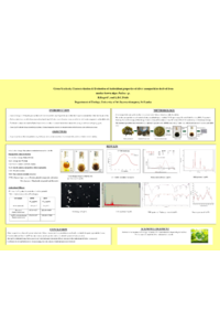
Padina sp., an edible brown macroalgae found along Sri Lanka's coast, is known for its bioactive compounds, eco-friendliness, and therapeutic benefits. These compounds can reduce metal ions to form nanoparticles, acting as stabilizing and capping agents. Silver nanoparticles, renowned for their conductivity, stability, and disease-treating potential, were synthesized in this study using Padina sp. The objectives were to synthesize and characterize the nanoparticles and evaluate their antioxidant activity. Silver nanoparticles were synthesized by mixing crude methanol extract of Padina sp. with silver nitrate. Characterization was conducted using Ultraviolet–Visible (UV-Vis) spectroscopy, Dynamic Light Scattering (DLS), Zeta potential analysis, Scanning Electron Microscopy (SEM), Energy-Dispersive X-ray (EDX) analysis, X-ray Diffraction (XRD), Fourier-Transform Infrared (FTIR) spectroscopy, and Raman spectroscopy. Antioxidant activity was assessed using the 2,2-Diphenyl-1-picrylhydrazyl (DPPH) assay. Statistical data analysis was performed using Minitab, with the results expressed as the mean ± standard deviation (SD), and paired two-sample t-tests were used to assess significant differences (p < 0.05). A visible color change from pale yellow to reddish-brown within 48 hours confirmed the formation of silver nanoparticles. UV-Vis spectroscopy showed a surface plasmon resonance peak at 420 nm, confirming the presence of nanoparticles. The DLS analysis revealed an average particle size of 73.19 nm, and the zeta potential obtained, -21.5 mV, indicated stability. The SEM images depicted spherical nanoparticles with smooth surfaces and no aggregation, while the EDX analysis confirmed 20% silver content by weight. The XRD analysis showed a face-centered cubic structure, indicating crystallinity. FTIR and Raman spectroscopy identified proteins, phenolic compounds, and amines as stabilizing agents, with polyphenolic compounds and flavonoids acting as reducing agents. In the DPPH assay, the antioxidant activity of silver nanoparticles (IC50 = 271.17 ± 3.99 μg/ml) was significantly higher than that of the crude Padina sp. extract (IC50 = 307.69 ± 9.33 μg/ml). These findings suggest that green-synthesized silver nanoparticles from Padina sp. offer a promising therapeutic strategy for diseases linked to oxidative stress.
Keywords
Silver nanoparticles
Characterization
Padina sp.
antioxidant
Poster
Ragavi -Poster - Sciforum 108008.pdf
Comparison of commonly used methods for efficient extraction and quantification of polyphenols
The antioxidant potential of Arbutus unedo tree berries from the Natural Park of Montesinho