Event submissions
Published
This submission belongs to the session S4. Immune System, Tumor Immunology and Autoimmune Disease of the event The 3rd International Electronic Conference on Biomedicines
Published date
13 May, 2025
Academic Editor
'%3e%3cpath%20d='M12.6657%2010.9104C12.4185%2010.325%2012.0599%209.79327%2011.6097%209.34482C11.1609%208.89508%2010.6293%208.5365%2010.0442%208.28888C10.0389%208.28626%2010.0337%208.28495%2010.0285%208.28233C10.8446%207.69279%2011.3752%206.73249%2011.3752%205.64904C11.3752%203.8542%209.92103%202.39999%208.1262%202.39999C6.33136%202.39999%204.87715%203.8542%204.87715%205.64904C4.87715%206.73249%205.40774%207.69279%206.22393%208.28364C6.21869%208.28626%206.21345%208.28757%206.20821%208.29019C5.62129%208.5378%205.09463%208.89284%204.64265%209.34613C4.1929%209.79493%203.83432%2010.3266%203.58671%2010.9117C3.34345%2011.4845%203.21226%2012.0987%203.20023%2012.7209C3.19988%2012.7349%203.20233%2012.7488%203.20744%2012.7619C3.21255%2012.7749%203.22022%2012.7867%203.22998%2012.7968C3.23975%2012.8068%203.25142%2012.8147%203.26431%2012.8202C3.2772%2012.8256%203.29105%2012.8284%203.30504%2012.8284H4.09109C4.14874%2012.8284%204.19459%2012.7825%204.1959%2012.7262C4.2221%2011.7148%204.62823%2010.7676%205.34617%2010.0497C6.08899%209.30683%207.0755%208.89808%208.1262%208.89808C9.17689%208.89808%2010.1634%209.30683%2010.9062%2010.0497C11.6242%2010.7676%2012.0303%2011.7148%2012.0565%2012.7262C12.0578%2012.7838%2012.1037%2012.8284%2012.1613%2012.8284H12.9474C12.9613%2012.8284%2012.9752%2012.8256%2012.9881%2012.8202C13.001%2012.8147%2013.0126%2012.8068%2013.0224%2012.7968C13.0322%2012.7867%2013.0398%2012.7749%2013.0449%2012.7619C13.0501%2012.7488%2013.0525%2012.7349%2013.0522%2012.7209C13.0391%2012.0947%2012.9094%2011.4855%2012.6657%2010.9104ZM8.1262%207.9024C7.52486%207.9024%206.9589%207.6679%206.53312%207.24211C6.10733%206.81633%205.87283%206.25037%205.87283%205.64904C5.87283%205.0477%206.10733%204.48174%206.53312%204.05596C6.9589%203.63018%207.52486%203.39567%208.1262%203.39567C8.72753%203.39567%209.29349%203.63018%209.71927%204.05596C10.1451%204.48174%2010.3796%205.0477%2010.3796%205.64904C10.3796%206.25037%2010.1451%206.81633%209.71927%207.24211C9.29349%207.6679%208.72753%207.9024%208.1262%207.9024Z'%20fill='%235D1EE1'/%3e%3c/g%3e%3c/svg%3e) Marianna Christodoulou
Marianna ChristodoulouCitation
Olga Pershina, Angelina Pakhomova, Olga Rybalkina, Ildar Uzyanbaev, Lena Kogai, Lubov Sandrikina, Elena Fedorova, Valentina Sedinova, Vadim Zhdanov, Pulmonary fibrosis and lung cancer: unity and difference of cellular processes, in Proceedings of The 3rd International Electronic Conference on Biomedicines, 12 May–15 May 2025, MDPI: Basel, Switzerland
Share
Email
Facebook
Twitter
LinkedIn
Pulmonary fibrosis and lung cancer: unity and difference of cellular processes
'%3e%3cpath%20d='M12.6647%2010.9104C12.4176%2010.325%2012.0589%209.7933%2011.6088%209.34485C11.16%208.89511%2010.6283%208.53653%2010.0432%208.28891C10.038%208.28629%2010.0327%208.28498%2010.0275%208.28236C10.8437%207.69282%2011.3743%206.73252%2011.3743%205.64907C11.3743%203.85423%209.92005%202.40002%208.12522%202.40002C6.33038%202.40002%204.87618%203.85423%204.87618%205.64907C4.87618%206.73252%205.40677%207.69282%206.22296%208.28367C6.21772%208.28629%206.21248%208.2876%206.20724%208.29022C5.62031%208.53783%205.09365%208.89287%204.64167%209.34616C4.19192%209.79496%203.83334%2010.3266%203.58573%2010.9117C3.34247%2011.4846%203.21128%2012.0987%203.19925%2012.721C3.1989%2012.735%203.20135%2012.7489%203.20646%2012.7619C3.21157%2012.7749%203.21924%2012.7868%203.22901%2012.7968C3.23877%2012.8068%203.25045%2012.8148%203.26334%2012.8202C3.27623%2012.8256%203.29007%2012.8284%203.30406%2012.8284H4.09012C4.14776%2012.8284%204.19362%2012.7825%204.19493%2012.7262C4.22113%2011.7148%204.62726%2010.7676%205.34519%2010.0497C6.08802%209.30686%207.07452%208.89811%208.12522%208.89811C9.17592%208.89811%2010.1624%209.30686%2010.9052%2010.0497C11.6232%2010.7676%2012.0293%2011.7148%2012.0555%2012.7262C12.0568%2012.7839%2012.1027%2012.8284%2012.1603%2012.8284H12.9464C12.9604%2012.8284%2012.9742%2012.8256%2012.9871%2012.8202C13%2012.8148%2013.0117%2012.8068%2013.0214%2012.7968C13.0312%2012.7868%2013.0389%2012.7749%2013.044%2012.7619C13.0491%2012.7489%2013.0515%2012.735%2013.0512%2012.721C13.0381%2012.0947%2012.9084%2011.4856%2012.6647%2010.9104ZM8.12522%207.90243C7.52388%207.90243%206.95792%207.66793%206.53214%207.24215C6.10636%206.81636%205.87185%206.2504%205.87185%205.64907C5.87185%205.04773%206.10636%204.48177%206.53214%204.05599C6.95792%203.63021%207.52388%203.3957%208.12522%203.3957C8.72655%203.3957%209.29252%203.63021%209.7183%204.05599C10.1441%204.48177%2010.3786%205.04773%2010.3786%205.64907C10.3786%206.2504%2010.1441%206.81636%209.7183%207.24215C9.29252%207.66793%208.72655%207.90243%208.12522%207.90243Z'%20fill='%235D1EE1'/%3e%3c/g%3e%3c/svg%3e)
Olga Pershina 1
Angelina Pakhomova 1,2
Olga Rybalkina 3
Ildar Uzyanbaev 1,4
Lena Kogai 1
Lubov Sandrikina 1,2
Elena Fedorova 1
Valentina Sedinova 1
Vadim Zhdanov 1
1. Laboratory of Regenerative Pharmacology, Goldberg ED Research Institute of Pharmacology and Regenerative Medicine, Tomsk National Research Medical Centre of the Russian Academy of Sciences, Tomsk, Russia
2. Normal Physiology Department, Siberian State Medical University, Tomsk, Russia
3. Goldberg ED Research Institute of Pharmacology and Regenerative Medicine, Tomsk National Research Medical Centre of the Russian Academy of Sciences, Tomsk, Russia
4. Siberian State Medical University, Tomsk, Russia
Abstract
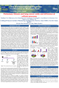
Despite the multiple identified common genetic, molecular, and cellular processes connecting pulmonary fibrosis (PF) and lung cancer (LC), there is still no clear answer about the contribution of each of them to the development of pathology. It is known that most cell populations involved in the development of PF are involved in LC. At the same time, there are differences that can be explained by the specificity of PF development or changes specific to LC. We evaluated the changes in the lung and blood at the modeling of PF, LC, PF+LC. To induce PF in male C57BL6 mice, we used bleomycin; on the 15th day after bleomycin administration, we modeled non-small cell LC by introducing Lewis lung carcinoma (LLC) cells. The material for this study was collected on the 28th day of the experiment and was analyzed by histology and cytometry. In the lung tissue of mice, against the background of PF and LC modeling, an increase in pathological changes in the lungs was observed compared to each pathological situation separately. Disorders of the lungs in the group of animals with PF and LC were based on increased migration from the blood to the lungs of populations of CD309+ endothelial cells, an increase in atypical/hybrid cells (CD45+CD326+), and cancer stem cell markers CD90, CD117, CD274, CD276, and EGFR in various combinations in the lung tissue. Biomarkers confirming tumor development against the background of pulmonary fibrosis are populations of cancer stem cells, atypical/hybrid cells, endothelial cells, hematopoietic stem cells, myeloid fibrocytes, and changes in the composition of CD4+ T-cells. Further clinical and experimental studies are needed to determine the role of these changes and to find biomarkers that predict the progression of pulmonary fibrosis and the risk of developing lung cancer.
Keywords
Pulmonary fibrosis
lung cancer
Lewis lung carcinoma
CD4+ T-cells
endothelial cells
cancer stem cells
Poster
PershinaO.pdf
Novel biomarkers for diagnosing prosthetic joint infection and chronic joint pain following total knee and hip arthroplasty: An exploratory study
Stimulation of the auricular vagus nerve in ST-Elevation Myocardial Infarction (STEMI)