Event submissions
Published
This submission belongs to the session S2. Novel Insights on the Mechanism of Action and/or Pathophysiology of Toxins of the event The 3rd International Online Conference on Toxins
Published date
08 Sep, 2025
Academic Editor
'%3e%3cpath%20d='M12.6657%2010.9104C12.4185%2010.325%2012.0599%209.79327%2011.6097%209.34482C11.1609%208.89508%2010.6293%208.5365%2010.0442%208.28888C10.0389%208.28626%2010.0337%208.28495%2010.0285%208.28233C10.8446%207.69279%2011.3752%206.73249%2011.3752%205.64904C11.3752%203.8542%209.92103%202.39999%208.1262%202.39999C6.33136%202.39999%204.87715%203.8542%204.87715%205.64904C4.87715%206.73249%205.40774%207.69279%206.22393%208.28364C6.21869%208.28626%206.21345%208.28757%206.20821%208.29019C5.62129%208.5378%205.09463%208.89284%204.64265%209.34613C4.1929%209.79493%203.83432%2010.3266%203.58671%2010.9117C3.34345%2011.4845%203.21226%2012.0987%203.20023%2012.7209C3.19988%2012.7349%203.20233%2012.7488%203.20744%2012.7619C3.21255%2012.7749%203.22022%2012.7867%203.22998%2012.7968C3.23975%2012.8068%203.25142%2012.8147%203.26431%2012.8202C3.2772%2012.8256%203.29105%2012.8284%203.30504%2012.8284H4.09109C4.14874%2012.8284%204.19459%2012.7825%204.1959%2012.7262C4.2221%2011.7148%204.62823%2010.7676%205.34617%2010.0497C6.08899%209.30683%207.0755%208.89808%208.1262%208.89808C9.17689%208.89808%2010.1634%209.30683%2010.9062%2010.0497C11.6242%2010.7676%2012.0303%2011.7148%2012.0565%2012.7262C12.0578%2012.7838%2012.1037%2012.8284%2012.1613%2012.8284H12.9474C12.9613%2012.8284%2012.9752%2012.8256%2012.9881%2012.8202C13.001%2012.8147%2013.0126%2012.8068%2013.0224%2012.7968C13.0322%2012.7867%2013.0398%2012.7749%2013.0449%2012.7619C13.0501%2012.7488%2013.0525%2012.7349%2013.0522%2012.7209C13.0391%2012.0947%2012.9094%2011.4855%2012.6657%2010.9104ZM8.1262%207.9024C7.52486%207.9024%206.9589%207.6679%206.53312%207.24211C6.10733%206.81633%205.87283%206.25037%205.87283%205.64904C5.87283%205.0477%206.10733%204.48174%206.53312%204.05596C6.9589%203.63018%207.52486%203.39567%208.1262%203.39567C8.72753%203.39567%209.29349%203.63018%209.71927%204.05596C10.1451%204.48174%2010.3796%205.0477%2010.3796%205.64904C10.3796%206.25037%2010.1451%206.81633%209.71927%207.24211C9.29349%207.6679%208.72753%207.9024%208.1262%207.9024Z'%20fill='%235D1EE1'/%3e%3c/g%3e%3c/svg%3e) Joseph Barbieri
Joseph BarbieriCitation
Miliausha Galimova, Oleg Pimenov, Ilia Teplov, Vladislav Starkov, Yuri Utkin, Effects of heterodimeric phospholipase A₂ from the venom of Vipera nikolskii on rat cardiomyocytes, in Proceedings of The 3rd International Online Conference on Toxins, 10 September–12 September 2025, MDPI: Basel, Switzerland
Share
Email
Facebook
Twitter
LinkedIn
Effects of heterodimeric phospholipase A2 from the venom of Vipera nikolskii on rat cardiomyocytes
'%3e%3cpath%20d='M12.6647%2010.9104C12.4176%2010.325%2012.0589%209.7933%2011.6088%209.34485C11.16%208.89511%2010.6283%208.53653%2010.0432%208.28891C10.038%208.28629%2010.0327%208.28498%2010.0275%208.28236C10.8437%207.69282%2011.3743%206.73252%2011.3743%205.64907C11.3743%203.85423%209.92005%202.40002%208.12522%202.40002C6.33038%202.40002%204.87618%203.85423%204.87618%205.64907C4.87618%206.73252%205.40677%207.69282%206.22296%208.28367C6.21772%208.28629%206.21248%208.2876%206.20724%208.29022C5.62031%208.53783%205.09365%208.89287%204.64167%209.34616C4.19192%209.79496%203.83334%2010.3266%203.58573%2010.9117C3.34247%2011.4846%203.21128%2012.0987%203.19925%2012.721C3.1989%2012.735%203.20135%2012.7489%203.20646%2012.7619C3.21157%2012.7749%203.21924%2012.7868%203.22901%2012.7968C3.23877%2012.8068%203.25045%2012.8148%203.26334%2012.8202C3.27623%2012.8256%203.29007%2012.8284%203.30406%2012.8284H4.09012C4.14776%2012.8284%204.19362%2012.7825%204.19493%2012.7262C4.22113%2011.7148%204.62726%2010.7676%205.34519%2010.0497C6.08802%209.30686%207.07452%208.89811%208.12522%208.89811C9.17592%208.89811%2010.1624%209.30686%2010.9052%2010.0497C11.6232%2010.7676%2012.0293%2011.7148%2012.0555%2012.7262C12.0568%2012.7839%2012.1027%2012.8284%2012.1603%2012.8284H12.9464C12.9604%2012.8284%2012.9742%2012.8256%2012.9871%2012.8202C13%2012.8148%2013.0117%2012.8068%2013.0214%2012.7968C13.0312%2012.7868%2013.0389%2012.7749%2013.044%2012.7619C13.0491%2012.7489%2013.0515%2012.735%2013.0512%2012.721C13.0381%2012.0947%2012.9084%2011.4856%2012.6647%2010.9104ZM8.12522%207.90243C7.52388%207.90243%206.95792%207.66793%206.53214%207.24215C6.10636%206.81636%205.87185%206.2504%205.87185%205.64907C5.87185%205.04773%206.10636%204.48177%206.53214%204.05599C6.95792%203.63021%207.52388%203.3957%208.12522%203.3957C8.72655%203.3957%209.29252%203.63021%209.7183%204.05599C10.1441%204.48177%2010.3786%205.04773%2010.3786%205.64907C10.3786%206.2504%2010.1441%206.81636%209.7183%207.24215C9.29252%207.66793%208.72655%207.90243%208.12522%207.90243Z'%20fill='%235D1EE1'/%3e%3c/g%3e%3c/svg%3e)
Miliausha Galimova 1
Oleg Pimenov 1
Ilia Teplov 1
Vladislav Starkov 2

Yuri Utkin 2
1. Institute of Theoretical and Experimental Biophysics, Russian Academy of Sciences, Pushchino 142290, Russia
2. Shemyakin-Ovchinnikov Institute of Bioorganic Chemistry, Russian Academy of Sciences, Moscow 117997, Russia
Abstract
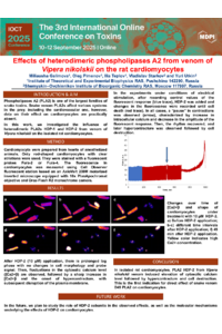
Introduction: Phospholipase A2 (PLA2) is one of the largest families of snake toxins. Snake venom PLA2s affect various systems in prey, including the cardiovascular one; however, data on their effect on cardiomyocytes are practically absent. In this work, we investigated the influence of heterodimeric PLA2s HDP-I and HDP-II from the venom of Vipera nikolskii on isolated rat cardiomyocytes.
Methods: Cardiomyocytes were prepared from the hearts of anesthetized animals. Only rod-shaped cardiomyocytes with clear striations were used. They were stained with a fluorescent probe, Fura-2 or Fura-4. The fluorescence in cardiomyocytes was measured using the Cell Observer fluorescent station based on an AxioVert 200M motorized inverted microscope equipped with a 10x PlanApochromat objective and Orca-Flash R2 monochrome camera.
Results: After HDP2 (10 μM) application, there was a prolonged lag phase with no changes in cell morphology or probe signal. Then, fluctuations in the cytosolic calcium level ([Ca2+]i) were observed, followed by a sharp increase in [Ca2+]i and the onset of hypercontracture, with subsequent disruption of the plasma membrane. In the experiments under conditions of electrical stimulation, after recording control values of the fluorescent response, HDP II was added and changes in the fluorescence were recorded until cell death. In all cases, a “pause” in contractions was observed, characterized by an increase in intracellular calcium and decrease in the amplitude of the fluorescent response. Then, the rhythm recovered, and later, hypercontracture was observed followed by cell destruction. The times to these events depended on the HDP II concentration and the efficiency of stimulation. No effects on cardiomyocytes were observed upon application of 1 µM HDP I.
Conclusions: In isolated rat cardiomyocytes, PLA2 HDP II from Vipera nikolskii venom induced an elevation in cytosolic calcium level followed by hypercontracture and cell destruction. This is the first indication for the direct effect of snake venom D49 PLA2 on cardiomyocytes.
Keywords
Phospholipase A2
cardiomyocyte
snake venom
fluorescence
hypercontracture
Poster
Poster-Utkin.pdf
Combined Detection of Tetrodotoxin and Saxitoxin in Seafood with a Multiplexed Electrochemical Biosensor
Epilepsy-associated mutation T226R in Kv1.1 subunit: properties of heterotetrameric Kv1.1(T226R)-Kv1.2 channel