Event submissions
Published

This submission belongs to the session c. Physical Sensors of the event International Electronic Conference on Sensors and Applications
Published date
02 Jun, 2014
Citation
Carlo Camerlingo, Fabrizia d'Apuzzo, V. Grassia, L. Perillo, Maria Lepore, Micro-Raman Spectroscopy for Assessment of Periodontal Disease Follow-Up, in Proceedings of International Electronic Conference on Sensors and Applications, 1 June–16 June 2014, MDPI: Basel, Switzerland, doi: 10.3390/ecsa-1-c003
Share
Email
Facebook
Twitter
LinkedIn
Micro-Raman Spectroscopy for Assessment of Periodontal Disease Follow-Up
'%3e%3cpath%20d='M12.6647%2010.9104C12.4176%2010.325%2012.0589%209.7933%2011.6088%209.34485C11.16%208.89511%2010.6283%208.53653%2010.0432%208.28891C10.038%208.28629%2010.0327%208.28498%2010.0275%208.28236C10.8437%207.69282%2011.3743%206.73252%2011.3743%205.64907C11.3743%203.85423%209.92005%202.40002%208.12522%202.40002C6.33038%202.40002%204.87618%203.85423%204.87618%205.64907C4.87618%206.73252%205.40677%207.69282%206.22296%208.28367C6.21772%208.28629%206.21248%208.2876%206.20724%208.29022C5.62031%208.53783%205.09365%208.89287%204.64167%209.34616C4.19192%209.79496%203.83334%2010.3266%203.58573%2010.9117C3.34247%2011.4846%203.21128%2012.0987%203.19925%2012.721C3.1989%2012.735%203.20135%2012.7489%203.20646%2012.7619C3.21157%2012.7749%203.21924%2012.7868%203.22901%2012.7968C3.23877%2012.8068%203.25045%2012.8148%203.26334%2012.8202C3.27623%2012.8256%203.29007%2012.8284%203.30406%2012.8284H4.09012C4.14776%2012.8284%204.19362%2012.7825%204.19493%2012.7262C4.22113%2011.7148%204.62726%2010.7676%205.34519%2010.0497C6.08802%209.30686%207.07452%208.89811%208.12522%208.89811C9.17592%208.89811%2010.1624%209.30686%2010.9052%2010.0497C11.6232%2010.7676%2012.0293%2011.7148%2012.0555%2012.7262C12.0568%2012.7839%2012.1027%2012.8284%2012.1603%2012.8284H12.9464C12.9604%2012.8284%2012.9742%2012.8256%2012.9871%2012.8202C13%2012.8148%2013.0117%2012.8068%2013.0214%2012.7968C13.0312%2012.7868%2013.0389%2012.7749%2013.044%2012.7619C13.0491%2012.7489%2013.0515%2012.735%2013.0512%2012.721C13.0381%2012.0947%2012.9084%2011.4856%2012.6647%2010.9104ZM8.12522%207.90243C7.52388%207.90243%206.95792%207.66793%206.53214%207.24215C6.10636%206.81636%205.87185%206.2504%205.87185%205.64907C5.87185%205.04773%206.10636%204.48177%206.53214%204.05599C6.95792%203.63021%207.52388%203.3957%208.12522%203.3957C8.72655%203.3957%209.29252%203.63021%209.7183%204.05599C10.1441%204.48177%2010.3786%205.04773%2010.3786%205.64907C10.3786%206.2504%2010.1441%206.81636%209.7183%207.24215C9.29252%207.66793%208.72655%207.90243%208.12522%207.90243Z'%20fill='%235D1EE1'/%3e%3c/g%3e%3c/svg%3e)
Carlo Camerlingo 1

Fabrizia d'Apuzzo 2
V. Grassia 2
L. Perillo 2

Maria Lepore 3
1. CNR-SPIN, Istituto superconduttori, materiali innovativi e dispositivi, via Campi Flegrei 34 , 80078 Pozzuoli, Italy;
2. Dipt. Multidisciplinare di Specialità Medico-Chirurgiche e Odontoiatriche, Seconda Università di Napoli, Napoli, Italy
3. Dipt. di Medicina Sperimentale, Università della Campania "L. Vanvitelli", Napoli, Italy
Abstract
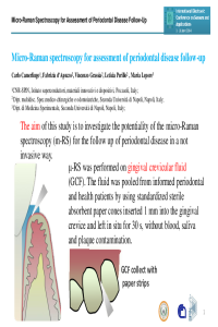
The aim of this study was to investigate the potentiality of the Micro-Raman spectroscopy in a follow up of the periodontal disease by performing a clinical not invasive sample collection method. Samples of gingival crevicular fluid (GCF) from informed periodontal and health patients were pooled. Specifically, the gingival crevicular fluid was collected with #30 standardized sterile absorbent paper cones inserted 1 mm into the gingival crevice and left in situ for 30 s [1]. The intra-oral clinical examinations were performed on one buccal site per maxillary central incisor. Each sample was tested by micro-Raman spectrometry. Raman spectra showed the presence of carotene in GCF periodontal patient's samples. With osteopotin, the carotene is expected to be correlates in crevicular fluid and salive with periodontal disease [2]. An automatic numerical data treatment based on wavelet algorithm was used in order to suppress the non-correlated signal, to subtract the background signal and to increase the quantitative readability of the Raman signal [3]. Micro-Raman spectroscopy is a very promising tool for medical applications, for its sensitivity to subtle changes in the chemical and structural characteristics of biological specimens [4]. [1] G. Perinetti, M. Paolantonio, B. Femminella, E. Serra, G. Spoto. G, J Periodontol 79 (2008) , 1200. [2] C.G. Sharma, A.R. Pradeep. J Periodont Res 42(2007) 450.[3] C. Camerlingo, F. Zenone, G.M. Gaeta, R. Riccio, M. Lepore. Meas Sci Technol 17 (2006) 298. [4] C. Camerlingo, F. Zenone, G. Perna, V. Capozzi, N. Cirillo, G.M. Gaeta, M. Lepore. . Sensors 8 (2008) 3656-64.
Keywords
Micro-Raman spectroscopy,
Manuscript
ECSA-1_Micro-Raman Spectroscopy_Camerlingo et al.pdf
Poster
ECSA-1_Micro-Raman Spectroscopy_Presentation_Camerlingo et al.pdf
A Novel MEMS based Surface Acoustic Wave Gas Sensor for Carbon Dioxide Detection in Hot-Process Areas
Analysis of Radio Wave Propagation for ISM 2.4GHz Wireless Sensor Networks in Inhomogeneous Vegetation Environments