Event submissions
Published
This submission belongs to the session S2. Dementia Spectrum Disorders: From Frontotemporal Degeneration to Parkinson’s and Alzheimer’s Disease of the event The 2nd International Electronic Conference on Medicine
Published date
06 Nov, 2025
Academic Editor
'%3e%3cpath%20d='M12.6657%2010.9104C12.4185%2010.325%2012.0599%209.79327%2011.6097%209.34482C11.1609%208.89508%2010.6293%208.5365%2010.0442%208.28888C10.0389%208.28626%2010.0337%208.28495%2010.0285%208.28233C10.8446%207.69279%2011.3752%206.73249%2011.3752%205.64904C11.3752%203.8542%209.92103%202.39999%208.1262%202.39999C6.33136%202.39999%204.87715%203.8542%204.87715%205.64904C4.87715%206.73249%205.40774%207.69279%206.22393%208.28364C6.21869%208.28626%206.21345%208.28757%206.20821%208.29019C5.62129%208.5378%205.09463%208.89284%204.64265%209.34613C4.1929%209.79493%203.83432%2010.3266%203.58671%2010.9117C3.34345%2011.4845%203.21226%2012.0987%203.20023%2012.7209C3.19988%2012.7349%203.20233%2012.7488%203.20744%2012.7619C3.21255%2012.7749%203.22022%2012.7867%203.22998%2012.7968C3.23975%2012.8068%203.25142%2012.8147%203.26431%2012.8202C3.2772%2012.8256%203.29105%2012.8284%203.30504%2012.8284H4.09109C4.14874%2012.8284%204.19459%2012.7825%204.1959%2012.7262C4.2221%2011.7148%204.62823%2010.7676%205.34617%2010.0497C6.08899%209.30683%207.0755%208.89808%208.1262%208.89808C9.17689%208.89808%2010.1634%209.30683%2010.9062%2010.0497C11.6242%2010.7676%2012.0303%2011.7148%2012.0565%2012.7262C12.0578%2012.7838%2012.1037%2012.8284%2012.1613%2012.8284H12.9474C12.9613%2012.8284%2012.9752%2012.8256%2012.9881%2012.8202C13.001%2012.8147%2013.0126%2012.8068%2013.0224%2012.7968C13.0322%2012.7867%2013.0398%2012.7749%2013.0449%2012.7619C13.0501%2012.7488%2013.0525%2012.7349%2013.0522%2012.7209C13.0391%2012.0947%2012.9094%2011.4855%2012.6657%2010.9104ZM8.1262%207.9024C7.52486%207.9024%206.9589%207.6679%206.53312%207.24211C6.10733%206.81633%205.87283%206.25037%205.87283%205.64904C5.87283%205.0477%206.10733%204.48174%206.53312%204.05596C6.9589%203.63018%207.52486%203.39567%208.1262%203.39567C8.72753%203.39567%209.29349%203.63018%209.71927%204.05596C10.1451%204.48174%2010.3796%205.0477%2010.3796%205.64904C10.3796%206.25037%2010.1451%206.81633%209.71927%207.24211C9.29349%207.6679%208.72753%207.9024%208.1262%207.9024Z'%20fill='%235D1EE1'/%3e%3c/g%3e%3c/svg%3e) Allison B. Reiss
Allison B. ReissCitation
Dimitra Sali, Dimitris Agiomyrgiannakis, Vassiliki Kamtsadeli, Maria Hatzopoulou, Evi Lykou, Niki Tsinia, Panagiotis Toulas, John Papatriantafyllou, THE USE OF VISUAL RATING SCALES IN THE DISCRIMINATION BETWEEN ALZHEIMER’S DISEASE AND DEPRESSION, in Proceedings of The 2nd International Electronic Conference on Medicine, 11 November–13 November 2025, MDPI: Basel, Switzerland
Share
Email
Facebook
Twitter
LinkedIn
THE USE OF VISUAL RATING SCALES IN THE DISCRIMINATION BETWEEN ALZHEIMER’S DISEASE AND DEPRESSION
'%3e%3cpath%20d='M12.6647%2010.9104C12.4176%2010.325%2012.0589%209.7933%2011.6088%209.34485C11.16%208.89511%2010.6283%208.53653%2010.0432%208.28891C10.038%208.28629%2010.0327%208.28498%2010.0275%208.28236C10.8437%207.69282%2011.3743%206.73252%2011.3743%205.64907C11.3743%203.85423%209.92005%202.40002%208.12522%202.40002C6.33038%202.40002%204.87618%203.85423%204.87618%205.64907C4.87618%206.73252%205.40677%207.69282%206.22296%208.28367C6.21772%208.28629%206.21248%208.2876%206.20724%208.29022C5.62031%208.53783%205.09365%208.89287%204.64167%209.34616C4.19192%209.79496%203.83334%2010.3266%203.58573%2010.9117C3.34247%2011.4846%203.21128%2012.0987%203.19925%2012.721C3.1989%2012.735%203.20135%2012.7489%203.20646%2012.7619C3.21157%2012.7749%203.21924%2012.7868%203.22901%2012.7968C3.23877%2012.8068%203.25045%2012.8148%203.26334%2012.8202C3.27623%2012.8256%203.29007%2012.8284%203.30406%2012.8284H4.09012C4.14776%2012.8284%204.19362%2012.7825%204.19493%2012.7262C4.22113%2011.7148%204.62726%2010.7676%205.34519%2010.0497C6.08802%209.30686%207.07452%208.89811%208.12522%208.89811C9.17592%208.89811%2010.1624%209.30686%2010.9052%2010.0497C11.6232%2010.7676%2012.0293%2011.7148%2012.0555%2012.7262C12.0568%2012.7839%2012.1027%2012.8284%2012.1603%2012.8284H12.9464C12.9604%2012.8284%2012.9742%2012.8256%2012.9871%2012.8202C13%2012.8148%2013.0117%2012.8068%2013.0214%2012.7968C13.0312%2012.7868%2013.0389%2012.7749%2013.044%2012.7619C13.0491%2012.7489%2013.0515%2012.735%2013.0512%2012.721C13.0381%2012.0947%2012.9084%2011.4856%2012.6647%2010.9104ZM8.12522%207.90243C7.52388%207.90243%206.95792%207.66793%206.53214%207.24215C6.10636%206.81636%205.87185%206.2504%205.87185%205.64907C5.87185%205.04773%206.10636%204.48177%206.53214%204.05599C6.95792%203.63021%207.52388%203.3957%208.12522%203.3957C8.72655%203.3957%209.29252%203.63021%209.7183%204.05599C10.1441%204.48177%2010.3786%205.04773%2010.3786%205.64907C10.3786%206.2504%2010.1441%206.81636%209.7183%207.24215C9.29252%207.66793%208.72655%207.90243%208.12522%207.90243Z'%20fill='%235D1EE1'/%3e%3c/g%3e%3c/svg%3e)
Dimitra Sali 1,2
Dimitris Agiomyrgiannakis 3
Vassiliki Kamtsadeli 3
Maria Hatzopoulou 3
Evi Lykou 3
Niki Tsinia 3
Panagiotis Toulas 4
John Papatriantafyllou 3
1. Athens Euroclinic General Hospital, Athanasiadou 7-9, Athens 115 21, Greece
2. Aiginiteio University Hospital, Vasilissis Sofias 72-74, Athens 115 28, Greece
3. Iasis-AMKE Geriatric Outpatient Clinic, Krimeas 73, Athens 165 62, Greece
4. Bioiatriki Diagnostic Centre, L.Kifisias 132, Athens 11526 , Greece
Abstract
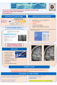
BACKGROUND: The discrimination of dementia and depression remains an issue of debate as they often co-exist in late life. Various neuroimaging techniques have been used in an attempt to differentiate between the two entities. Visual rating seems to offer a quick and reliable tool for diagnosis. We performed a retrospective study to assess the potential role of the visual rating method to assist in the differential diagnosis of AD versus depression.
METHODS: A sample of memory clinic patients fulfilling clinical criteria of either AD (n = 113) or depression (n = 139) was recruited. Depressive symptoms were defined as a score of 10 or higher on the GDS. Patients with AD were at a mild stage of disease (MMSE>23). Based on previous studies, 13 regions of interest were selected in both hemispheres, including mainly frontal, temporal and hippocampal areas. The regions were validated by two raters by using a visual scale with four stages of atrophy (0 to 4). Data were calculated using the binary logistic regression model.
RESULTS: Based on visual assessment, patients with AD showed more atrophy in the right anterior cingulate cortex and in temporal, limbic and hippocampal regions in comparison to patients with depression.
CONCLUSION: Visual rating scales offer a useful method to distinguish between AD and deperession in clinical practice.
Keywords
alzeimer disease
dementia
depression
Poster
POSTER IECM 2025.pdf
Hospital Morbidity due to Parkinson's Disease in Brazilian Regions between 2020 and 2024
Trends in hospitalizations for Alzheimer’s disease in the Brazilian unified health system, 2016–2025: age group analysis and evidence of early-onset cases