Event submissions
Published
This submission belongs to the session S4. Gels in Medicine, Regenerative Medicine, Pharmacy, and Personal Care Products of the event The 1st International Online Conference on Gels
Published date
28 Nov, 2025
Academic Editor
'%3e%3cpath%20d='M12.6657%2010.9104C12.4185%2010.325%2012.0599%209.79327%2011.6097%209.34482C11.1609%208.89508%2010.6293%208.5365%2010.0442%208.28888C10.0389%208.28626%2010.0337%208.28495%2010.0285%208.28233C10.8446%207.69279%2011.3752%206.73249%2011.3752%205.64904C11.3752%203.8542%209.92103%202.39999%208.1262%202.39999C6.33136%202.39999%204.87715%203.8542%204.87715%205.64904C4.87715%206.73249%205.40774%207.69279%206.22393%208.28364C6.21869%208.28626%206.21345%208.28757%206.20821%208.29019C5.62129%208.5378%205.09463%208.89284%204.64265%209.34613C4.1929%209.79493%203.83432%2010.3266%203.58671%2010.9117C3.34345%2011.4845%203.21226%2012.0987%203.20023%2012.7209C3.19988%2012.7349%203.20233%2012.7488%203.20744%2012.7619C3.21255%2012.7749%203.22022%2012.7867%203.22998%2012.7968C3.23975%2012.8068%203.25142%2012.8147%203.26431%2012.8202C3.2772%2012.8256%203.29105%2012.8284%203.30504%2012.8284H4.09109C4.14874%2012.8284%204.19459%2012.7825%204.1959%2012.7262C4.2221%2011.7148%204.62823%2010.7676%205.34617%2010.0497C6.08899%209.30683%207.0755%208.89808%208.1262%208.89808C9.17689%208.89808%2010.1634%209.30683%2010.9062%2010.0497C11.6242%2010.7676%2012.0303%2011.7148%2012.0565%2012.7262C12.0578%2012.7838%2012.1037%2012.8284%2012.1613%2012.8284H12.9474C12.9613%2012.8284%2012.9752%2012.8256%2012.9881%2012.8202C13.001%2012.8147%2013.0126%2012.8068%2013.0224%2012.7968C13.0322%2012.7867%2013.0398%2012.7749%2013.0449%2012.7619C13.0501%2012.7488%2013.0525%2012.7349%2013.0522%2012.7209C13.0391%2012.0947%2012.9094%2011.4855%2012.6657%2010.9104ZM8.1262%207.9024C7.52486%207.9024%206.9589%207.6679%206.53312%207.24211C6.10733%206.81633%205.87283%206.25037%205.87283%205.64904C5.87283%205.0477%206.10733%204.48174%206.53312%204.05596C6.9589%203.63018%207.52486%203.39567%208.1262%203.39567C8.72753%203.39567%209.29349%203.63018%209.71927%204.05596C10.1451%204.48174%2010.3796%205.0477%2010.3796%205.64904C10.3796%206.25037%2010.1451%206.81633%209.71927%207.24211C9.29349%207.6679%208.72753%207.9024%208.1262%207.9024Z'%20fill='%235D1EE1'/%3e%3c/g%3e%3c/svg%3e) Dirk Kuckling
Dirk KucklingCitation
Hongji Zhang, Fengjie Zhang, Chao Wan, Boosting engineered cartilage formation with recombinant spider silk, in Proceedings of The 1st International Online Conference on Gels, 3 December–5 December 2025, MDPI: Basel, Switzerland
Share
Email
Facebook
Twitter
LinkedIn
Boosting engineered cartilage formation with recombinant spider silk
'%3e%3cpath%20d='M12.6647%2010.9104C12.4176%2010.325%2012.0589%209.7933%2011.6088%209.34485C11.16%208.89511%2010.6283%208.53653%2010.0432%208.28891C10.038%208.28629%2010.0327%208.28498%2010.0275%208.28236C10.8437%207.69282%2011.3743%206.73252%2011.3743%205.64907C11.3743%203.85423%209.92005%202.40002%208.12522%202.40002C6.33038%202.40002%204.87618%203.85423%204.87618%205.64907C4.87618%206.73252%205.40677%207.69282%206.22296%208.28367C6.21772%208.28629%206.21248%208.2876%206.20724%208.29022C5.62031%208.53783%205.09365%208.89287%204.64167%209.34616C4.19192%209.79496%203.83334%2010.3266%203.58573%2010.9117C3.34247%2011.4846%203.21128%2012.0987%203.19925%2012.721C3.1989%2012.735%203.20135%2012.7489%203.20646%2012.7619C3.21157%2012.7749%203.21924%2012.7868%203.22901%2012.7968C3.23877%2012.8068%203.25045%2012.8148%203.26334%2012.8202C3.27623%2012.8256%203.29007%2012.8284%203.30406%2012.8284H4.09012C4.14776%2012.8284%204.19362%2012.7825%204.19493%2012.7262C4.22113%2011.7148%204.62726%2010.7676%205.34519%2010.0497C6.08802%209.30686%207.07452%208.89811%208.12522%208.89811C9.17592%208.89811%2010.1624%209.30686%2010.9052%2010.0497C11.6232%2010.7676%2012.0293%2011.7148%2012.0555%2012.7262C12.0568%2012.7839%2012.1027%2012.8284%2012.1603%2012.8284H12.9464C12.9604%2012.8284%2012.9742%2012.8256%2012.9871%2012.8202C13%2012.8148%2013.0117%2012.8068%2013.0214%2012.7968C13.0312%2012.7868%2013.0389%2012.7749%2013.044%2012.7619C13.0491%2012.7489%2013.0515%2012.735%2013.0512%2012.721C13.0381%2012.0947%2012.9084%2011.4856%2012.6647%2010.9104ZM8.12522%207.90243C7.52388%207.90243%206.95792%207.66793%206.53214%207.24215C6.10636%206.81636%205.87185%206.2504%205.87185%205.64907C5.87185%205.04773%206.10636%204.48177%206.53214%204.05599C6.95792%203.63021%207.52388%203.3957%208.12522%203.3957C8.72655%203.3957%209.29252%203.63021%209.7183%204.05599C10.1441%204.48177%2010.3786%205.04773%2010.3786%205.64907C10.3786%206.2504%2010.1441%206.81636%209.7183%207.24215C9.29252%207.66793%208.72655%207.90243%208.12522%207.90243Z'%20fill='%235D1EE1'/%3e%3c/g%3e%3c/svg%3e)
Hongji Zhang 1,2
Fengjie Zhang 1,2
Chao Wan 1,2
1. Center for Neuromusculoskeletal Restorative Medicine, Hong Kong Science Park, Hong Kong SAR, China
2. Key Laboratory of Regenerative Medicine, Ministry of Education (Shenzhen Base), School of Biomedical Sciences Core Laboratory, Shenzhen Research Institute, The Chinese University of Hong Kong, Shenzhen 518057, China
Abstract
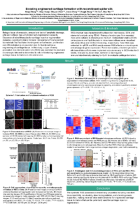
Introduction: Articular cartilage, being avascular and aneural and lacking lymphatic drainage, exhibits minimal intrinsic capacity for self-repair. The development of effective therapies for cartilage regeneration therefore remains an unmet clinical need. A critical step toward functional cartilage tissue engineering is the design of biomaterials that promote chondrocyte adhesion, proliferation and differentiation. In this study, we examined a novel protein-based silk material for its ability to modulate chondrocyte behavior and support engineered cartilage formation both in vitro and in vivo.
Methods: Primary chondrocytes were isolated from neonatal mice and cultured in chondrogenic medium. In vitro, chondrocytes were co-cultured with a type of recombinant spider silk. Extracellular matrix (ECM) proteoglycan deposition was assessed by Alcian blue staining, while protein expression was evaluated by Western blot. Total RNA was extracted from micromass cultures, and chondrogenic markers (Sox9, Col2α1, and aggrecan) were quantified using real-time PCR. For in vivo studies, chondrocyte-laden silk bioscaffolds were implanted subcutaneously into SCID mice for 3 and 6 weeks. Engineered cartilage formation was analyzed histologically using Alcian blue and Safranin O staining.
Results: The protein-based silk material created a favorable 3D microenvironment that promoted strong chondrocyte adhesion and enhanced chondrogenic differentiation. In vitro, chondrocytes maintained high viability on the silk material, with conditioned cultures showing elevated cartilage-specific glycosaminoglycan accumulation and the upregulated expression of chondrogenic genes. A high cell density was observed at the material interface, indicating robust cell–material interactions. In vivo, silk-based chondrocyte bioscaffolds supported cartilage tissue formation after implantation in SCID mice, as confirmed by histological analysis.
Conclusion: The synthetic protein-based silk material exhibits excellent biocompatibility, non-toxicity, controlled biodegradability and strong chondrogenesis-supportive properties. These findings highlight its potential as a promising scaffold for engineered cartilage tissue and as a candidate for future cartilage repair and regeneration therapies.
Keywords
tissue engineering
cartilage regeneration
biomaterial
recombinant protein
Poster
IOCG-Boosting engineered cartilage formation with recombinant spider silk.pdf
Quantum-Programmed Nanogels for Ultra-Precise Cancer Therapy
Impact of Storage on the Structure Stability of Starch-Based HPP Hydrogels Loaded with Natural Extracts