Event submissions
Published
This submission belongs to the session S1. Applied Biosciences and Bioengineering of the event The 6th International Electronic Conference on Applied Sciences
Published date
03 Dec, 2025
Academic Editor
'%3e%3cpath%20d='M12.6657%2010.9104C12.4185%2010.325%2012.0599%209.79327%2011.6097%209.34482C11.1609%208.89508%2010.6293%208.5365%2010.0442%208.28888C10.0389%208.28626%2010.0337%208.28495%2010.0285%208.28233C10.8446%207.69279%2011.3752%206.73249%2011.3752%205.64904C11.3752%203.8542%209.92103%202.39999%208.1262%202.39999C6.33136%202.39999%204.87715%203.8542%204.87715%205.64904C4.87715%206.73249%205.40774%207.69279%206.22393%208.28364C6.21869%208.28626%206.21345%208.28757%206.20821%208.29019C5.62129%208.5378%205.09463%208.89284%204.64265%209.34613C4.1929%209.79493%203.83432%2010.3266%203.58671%2010.9117C3.34345%2011.4845%203.21226%2012.0987%203.20023%2012.7209C3.19988%2012.7349%203.20233%2012.7488%203.20744%2012.7619C3.21255%2012.7749%203.22022%2012.7867%203.22998%2012.7968C3.23975%2012.8068%203.25142%2012.8147%203.26431%2012.8202C3.2772%2012.8256%203.29105%2012.8284%203.30504%2012.8284H4.09109C4.14874%2012.8284%204.19459%2012.7825%204.1959%2012.7262C4.2221%2011.7148%204.62823%2010.7676%205.34617%2010.0497C6.08899%209.30683%207.0755%208.89808%208.1262%208.89808C9.17689%208.89808%2010.1634%209.30683%2010.9062%2010.0497C11.6242%2010.7676%2012.0303%2011.7148%2012.0565%2012.7262C12.0578%2012.7838%2012.1037%2012.8284%2012.1613%2012.8284H12.9474C12.9613%2012.8284%2012.9752%2012.8256%2012.9881%2012.8202C13.001%2012.8147%2013.0126%2012.8068%2013.0224%2012.7968C13.0322%2012.7867%2013.0398%2012.7749%2013.0449%2012.7619C13.0501%2012.7488%2013.0525%2012.7349%2013.0522%2012.7209C13.0391%2012.0947%2012.9094%2011.4855%2012.6657%2010.9104ZM8.1262%207.9024C7.52486%207.9024%206.9589%207.6679%206.53312%207.24211C6.10733%206.81633%205.87283%206.25037%205.87283%205.64904C5.87283%205.0477%206.10733%204.48174%206.53312%204.05596C6.9589%203.63018%207.52486%203.39567%208.1262%203.39567C8.72753%203.39567%209.29349%203.63018%209.71927%204.05596C10.1451%204.48174%2010.3796%205.0477%2010.3796%205.64904C10.3796%206.25037%2010.1451%206.81633%209.71927%207.24211C9.29349%207.6679%208.72753%207.9024%208.1262%207.9024Z'%20fill='%235D1EE1'/%3e%3c/g%3e%3c/svg%3e) Roger Narayan
Roger NarayanCitation
Sergei V. Lepeshkevich, Igor V. Sazanovich, Marina V. Parkhats, Determination of Carbon Monoxide Dynamics in Hemoglobin Subunits Using Singular Value Decomposition and Maximum Entropy Method, in Proceedings of The 6th International Electronic Conference on Applied Sciences, 9 December–11 December 2025, MDPI: Basel, Switzerland
Share
Email
Facebook
Twitter
LinkedIn
Determination of Carbon Monoxide Dynamics in Hemoglobin Subunits Using Singular Value Decomposition and Maximum Entropy Method
'%3e%3cpath%20d='M12.6647%2010.9104C12.4176%2010.325%2012.0589%209.7933%2011.6088%209.34485C11.16%208.89511%2010.6283%208.53653%2010.0432%208.28891C10.038%208.28629%2010.0327%208.28498%2010.0275%208.28236C10.8437%207.69282%2011.3743%206.73252%2011.3743%205.64907C11.3743%203.85423%209.92005%202.40002%208.12522%202.40002C6.33038%202.40002%204.87618%203.85423%204.87618%205.64907C4.87618%206.73252%205.40677%207.69282%206.22296%208.28367C6.21772%208.28629%206.21248%208.2876%206.20724%208.29022C5.62031%208.53783%205.09365%208.89287%204.64167%209.34616C4.19192%209.79496%203.83334%2010.3266%203.58573%2010.9117C3.34247%2011.4846%203.21128%2012.0987%203.19925%2012.721C3.1989%2012.735%203.20135%2012.7489%203.20646%2012.7619C3.21157%2012.7749%203.21924%2012.7868%203.22901%2012.7968C3.23877%2012.8068%203.25045%2012.8148%203.26334%2012.8202C3.27623%2012.8256%203.29007%2012.8284%203.30406%2012.8284H4.09012C4.14776%2012.8284%204.19362%2012.7825%204.19493%2012.7262C4.22113%2011.7148%204.62726%2010.7676%205.34519%2010.0497C6.08802%209.30686%207.07452%208.89811%208.12522%208.89811C9.17592%208.89811%2010.1624%209.30686%2010.9052%2010.0497C11.6232%2010.7676%2012.0293%2011.7148%2012.0555%2012.7262C12.0568%2012.7839%2012.1027%2012.8284%2012.1603%2012.8284H12.9464C12.9604%2012.8284%2012.9742%2012.8256%2012.9871%2012.8202C13%2012.8148%2013.0117%2012.8068%2013.0214%2012.7968C13.0312%2012.7868%2013.0389%2012.7749%2013.044%2012.7619C13.0491%2012.7489%2013.0515%2012.735%2013.0512%2012.721C13.0381%2012.0947%2012.9084%2011.4856%2012.6647%2010.9104ZM8.12522%207.90243C7.52388%207.90243%206.95792%207.66793%206.53214%207.24215C6.10636%206.81636%205.87185%206.2504%205.87185%205.64907C5.87185%205.04773%206.10636%204.48177%206.53214%204.05599C6.95792%203.63021%207.52388%203.3957%208.12522%203.3957C8.72655%203.3957%209.29252%203.63021%209.7183%204.05599C10.1441%204.48177%2010.3786%205.04773%2010.3786%205.64907C10.3786%206.2504%2010.1441%206.81636%209.7183%207.24215C9.29252%207.66793%208.72655%207.90243%208.12522%207.90243Z'%20fill='%235D1EE1'/%3e%3c/g%3e%3c/svg%3e)
Sergei V. Lepeshkevich 1
Igor V. Sazanovich 2
Marina V. Parkhats 1
1. B.I. Stepanov Institute of Physics, National Academy of Sciences of Belarus, 68 Nezavisimosti Ave, Minsk 220072, Belarus.
2. Central Laser Facility, Research Complex at Harwell, STFC Rutherford Appleton Laboratory, Harwell Campus, OX11 0QX, UK.
Abstract
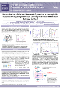
Human hemoglobin is a tetramer consisting of two α and two β subunits. Each subunit contains one identical ferrous heme group that can reversibly bind one ligand such as carbon monoxide (CO). Determining CO dynamics in hemoglobin subunits is essential for gaining insight into the transport of small molecules in physiological systems. Here, we use picosecond to millisecond transient mid-infrared (mid-IR) spectroscopy to study the photoinduced dynamics of CO in isolated hemoglobin subunits. Photoinduced absorption changes of the isolated carbonmonoxy hemoglobin subunits were measured after photoexcitation at 543 nm into the Q bands of the heme moiety. Time-resolved spectra in the mid-IR region were measured on the ULTRA apparatus at the Central Laser Facility (Didcot, UK). All the experiments were performed in 50 mM Tris buffer, pD 8.2, at 19°C.
The time evolution of the vibrational spectra of the coordinated and as photodissociated CO molecules was monitored in the spectral range between 1900 and 2180 cm-1. The mid-IR spectrum of the liganded subunits shows discrete CO stretch bands, denoted A0 (~1,968 cm–1) and A1 (~1,950 cm–1). The distinct stretch bands for CO photolyzed and temporarily trapped in the protein matrix are detected in the region of 2,090–2,160 cm–1. The measured transient mid-IR spectra were analyzed using singular value decomposition and maximum entropy method analysis. We succeeded in following the evolution of CO in hemoglobin subunits. The kinetic model, describing both the photodissociation and subsequent rebinding of CO, is introduced and discussed.
Keywords
carbon monoxide
hemoglobin
mid-infrared spectroscopy
dissociation
Poster
Lepeshkevich et al (ASEC 2025).pdf
Kinetic study of Diclofenac removal on Biocomposite microcapsules in aqueous systems
Next-Day Forest Fire Risk Prediction Using Machine Learning and Multimodal Satellite Data