Event submissions
Published
This submission belongs to the session S3. Computing and Artificial Intelligence of the event The 6th International Electronic Conference on Applied Sciences
Published date
03 Dec, 2025
Academic Editor
'%3e%3cpath%20d='M12.6657%2010.9104C12.4185%2010.325%2012.0599%209.79327%2011.6097%209.34482C11.1609%208.89508%2010.6293%208.5365%2010.0442%208.28888C10.0389%208.28626%2010.0337%208.28495%2010.0285%208.28233C10.8446%207.69279%2011.3752%206.73249%2011.3752%205.64904C11.3752%203.8542%209.92103%202.39999%208.1262%202.39999C6.33136%202.39999%204.87715%203.8542%204.87715%205.64904C4.87715%206.73249%205.40774%207.69279%206.22393%208.28364C6.21869%208.28626%206.21345%208.28757%206.20821%208.29019C5.62129%208.5378%205.09463%208.89284%204.64265%209.34613C4.1929%209.79493%203.83432%2010.3266%203.58671%2010.9117C3.34345%2011.4845%203.21226%2012.0987%203.20023%2012.7209C3.19988%2012.7349%203.20233%2012.7488%203.20744%2012.7619C3.21255%2012.7749%203.22022%2012.7867%203.22998%2012.7968C3.23975%2012.8068%203.25142%2012.8147%203.26431%2012.8202C3.2772%2012.8256%203.29105%2012.8284%203.30504%2012.8284H4.09109C4.14874%2012.8284%204.19459%2012.7825%204.1959%2012.7262C4.2221%2011.7148%204.62823%2010.7676%205.34617%2010.0497C6.08899%209.30683%207.0755%208.89808%208.1262%208.89808C9.17689%208.89808%2010.1634%209.30683%2010.9062%2010.0497C11.6242%2010.7676%2012.0303%2011.7148%2012.0565%2012.7262C12.0578%2012.7838%2012.1037%2012.8284%2012.1613%2012.8284H12.9474C12.9613%2012.8284%2012.9752%2012.8256%2012.9881%2012.8202C13.001%2012.8147%2013.0126%2012.8068%2013.0224%2012.7968C13.0322%2012.7867%2013.0398%2012.7749%2013.0449%2012.7619C13.0501%2012.7488%2013.0525%2012.7349%2013.0522%2012.7209C13.0391%2012.0947%2012.9094%2011.4855%2012.6657%2010.9104ZM8.1262%207.9024C7.52486%207.9024%206.9589%207.6679%206.53312%207.24211C6.10733%206.81633%205.87283%206.25037%205.87283%205.64904C5.87283%205.0477%206.10733%204.48174%206.53312%204.05596C6.9589%203.63018%207.52486%203.39567%208.1262%203.39567C8.72753%203.39567%209.29349%203.63018%209.71927%204.05596C10.1451%204.48174%2010.3796%205.0477%2010.3796%205.64904C10.3796%206.25037%2010.1451%206.81633%209.71927%207.24211C9.29349%207.6679%208.72753%207.9024%208.1262%207.9024Z'%20fill='%235D1EE1'/%3e%3c/g%3e%3c/svg%3e) Lucia Billeci
Lucia BilleciCitation
Nadenlla Rajamohan Reddy, G. Muneeswari, Segmentation of an Atypical Teratoid Rhabdoid Tumor Using UNet+ Fork with ResNext and ResNet for Improved MRI Analysis, in Proceedings of The 6th International Electronic Conference on Applied Sciences, 9 December–11 December 2025, MDPI: Basel, Switzerland
Share
Email
Facebook
Twitter
LinkedIn
Segmentation of an Atypical Teratoid Rhabdoid Tumor Using UNet+ Fork with ResNext and ResNet for Improved MRI Analysis
'%3e%3cpath%20d='M12.6647%2010.9104C12.4176%2010.325%2012.0589%209.7933%2011.6088%209.34485C11.16%208.89511%2010.6283%208.53653%2010.0432%208.28891C10.038%208.28629%2010.0327%208.28498%2010.0275%208.28236C10.8437%207.69282%2011.3743%206.73252%2011.3743%205.64907C11.3743%203.85423%209.92005%202.40002%208.12522%202.40002C6.33038%202.40002%204.87618%203.85423%204.87618%205.64907C4.87618%206.73252%205.40677%207.69282%206.22296%208.28367C6.21772%208.28629%206.21248%208.2876%206.20724%208.29022C5.62031%208.53783%205.09365%208.89287%204.64167%209.34616C4.19192%209.79496%203.83334%2010.3266%203.58573%2010.9117C3.34247%2011.4846%203.21128%2012.0987%203.19925%2012.721C3.1989%2012.735%203.20135%2012.7489%203.20646%2012.7619C3.21157%2012.7749%203.21924%2012.7868%203.22901%2012.7968C3.23877%2012.8068%203.25045%2012.8148%203.26334%2012.8202C3.27623%2012.8256%203.29007%2012.8284%203.30406%2012.8284H4.09012C4.14776%2012.8284%204.19362%2012.7825%204.19493%2012.7262C4.22113%2011.7148%204.62726%2010.7676%205.34519%2010.0497C6.08802%209.30686%207.07452%208.89811%208.12522%208.89811C9.17592%208.89811%2010.1624%209.30686%2010.9052%2010.0497C11.6232%2010.7676%2012.0293%2011.7148%2012.0555%2012.7262C12.0568%2012.7839%2012.1027%2012.8284%2012.1603%2012.8284H12.9464C12.9604%2012.8284%2012.9742%2012.8256%2012.9871%2012.8202C13%2012.8148%2013.0117%2012.8068%2013.0214%2012.7968C13.0312%2012.7868%2013.0389%2012.7749%2013.044%2012.7619C13.0491%2012.7489%2013.0515%2012.735%2013.0512%2012.721C13.0381%2012.0947%2012.9084%2011.4856%2012.6647%2010.9104ZM8.12522%207.90243C7.52388%207.90243%206.95792%207.66793%206.53214%207.24215C6.10636%206.81636%205.87185%206.2504%205.87185%205.64907C5.87185%205.04773%206.10636%204.48177%206.53214%204.05599C6.95792%203.63021%207.52388%203.3957%208.12522%203.3957C8.72655%203.3957%209.29252%203.63021%209.7183%204.05599C10.1441%204.48177%2010.3786%205.04773%2010.3786%205.64907C10.3786%206.2504%2010.1441%206.81636%209.7183%207.24215C9.29252%207.66793%208.72655%207.90243%208.12522%207.90243Z'%20fill='%235D1EE1'/%3e%3c/g%3e%3c/svg%3e)
Nadenlla Rajamohan Reddy 1
G. Muneeswari 1
1. School of Computer Science and Engineering, VIT-AP University, Amaravati, Andhra Pradesh-522237, India
Abstract
Atypical Teratoid Rhabdoid Tumor (ATRT) is a highly aggressive pediatric brain tumor, which poses substantial clinical challenges due to its rapid proliferation and complex morphological diversity. Precise segmentation of ATRT in Magnetic Resonance Imaging (MRI) is essential for accurate diagnosis, effective treatment planning, and thorough outcome evaluation. However, manual segmentation is time-consuming and susceptible to errors, particularly given the tumor's intricate structure. To overcome these limitations, deep learning-based automated segmentation techniques have attracted significant attention, with UNet architectures emerging as a leading solution in medical image analysis. This study introduces an advanced segmentation approach, utilizing a Fork of the Tumor-Segmentation-UNet+ model integrated with residual networks, namely ResNext and ResNet. These architectures enhance the model's ability to delineate complex tumor boundaries and account for the high degree of heterogeneity seen in ATRT. The inclusion of residual blocks from ResNext and ResNet facilitates more efficient feature extraction while also mitigating common issues in deep neural networks, such as vanishing gradients. The proposed model was trained and validated on a dataset comprising ATRT-specific MRI scans and compared against conventional segmentation approaches. Performance metrics, including the Dice coefficient, Intersection over Union (IoU), and sensitivity, were used to measure segmentation accuracy. The results indicate that the UNet+ model enhanced with ResNext and ResNet significantly outperforms standard UNet configurations, delivering more accurate and reliable segmentation of ATRT tumors.
Keywords
Atypical Teratoid Rhabdoid Tumor
MRI
Segmentation
Deep Learning
Residual Network
UNet+
and ResNext.
Poster
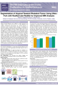
ASEC 2025 Poster Presentation.pdf
Classification of chemical coating quality in soybeans using convolutional neural networks
Machine Learning-Based Prognostic Modeling of Thyroid Cancer Recurrence