Event submissions
Published
This submission belongs to the session S5. Generative AI in Clinical Practice—Evidence-Based Evaluation of Diagnostic and Therapeutic Applications of the event The 1st International Online Conference on Healthcare
Published date
20 Mar, 2026
Academic Editor
'%3e%3cpath%20d='M12.6657%2010.9104C12.4185%2010.325%2012.0599%209.79327%2011.6097%209.34482C11.1609%208.89508%2010.6293%208.5365%2010.0442%208.28888C10.0389%208.28626%2010.0337%208.28495%2010.0285%208.28233C10.8446%207.69279%2011.3752%206.73249%2011.3752%205.64904C11.3752%203.8542%209.92103%202.39999%208.1262%202.39999C6.33136%202.39999%204.87715%203.8542%204.87715%205.64904C4.87715%206.73249%205.40774%207.69279%206.22393%208.28364C6.21869%208.28626%206.21345%208.28757%206.20821%208.29019C5.62129%208.5378%205.09463%208.89284%204.64265%209.34613C4.1929%209.79493%203.83432%2010.3266%203.58671%2010.9117C3.34345%2011.4845%203.21226%2012.0987%203.20023%2012.7209C3.19988%2012.7349%203.20233%2012.7488%203.20744%2012.7619C3.21255%2012.7749%203.22022%2012.7867%203.22998%2012.7968C3.23975%2012.8068%203.25142%2012.8147%203.26431%2012.8202C3.2772%2012.8256%203.29105%2012.8284%203.30504%2012.8284H4.09109C4.14874%2012.8284%204.19459%2012.7825%204.1959%2012.7262C4.2221%2011.7148%204.62823%2010.7676%205.34617%2010.0497C6.08899%209.30683%207.0755%208.89808%208.1262%208.89808C9.17689%208.89808%2010.1634%209.30683%2010.9062%2010.0497C11.6242%2010.7676%2012.0303%2011.7148%2012.0565%2012.7262C12.0578%2012.7838%2012.1037%2012.8284%2012.1613%2012.8284H12.9474C12.9613%2012.8284%2012.9752%2012.8256%2012.9881%2012.8202C13.001%2012.8147%2013.0126%2012.8068%2013.0224%2012.7968C13.0322%2012.7867%2013.0398%2012.7749%2013.0449%2012.7619C13.0501%2012.7488%2013.0525%2012.7349%2013.0522%2012.7209C13.0391%2012.0947%2012.9094%2011.4855%2012.6657%2010.9104ZM8.1262%207.9024C7.52486%207.9024%206.9589%207.6679%206.53312%207.24211C6.10733%206.81633%205.87283%206.25037%205.87283%205.64904C5.87283%205.0477%206.10733%204.48174%206.53312%204.05596C6.9589%203.63018%207.52486%203.39567%208.1262%203.39567C8.72753%203.39567%209.29349%203.63018%209.71927%204.05596C10.1451%204.48174%2010.3796%205.0477%2010.3796%205.64904C10.3796%206.25037%2010.1451%206.81633%209.71927%207.24211C9.29349%207.6679%208.72753%207.9024%208.1262%207.9024Z'%20fill='%235D1EE1'/%3e%3c/g%3e%3c/svg%3e) Lorraine Evangelista
Lorraine EvangelistaCitation
Oliwia Andrzejewska, Patrycja Chruniak, Mateusz Fornalski, Piotr Wójcik, Cezary Borysiuk, Michał Woś, Explainable AI for Detection of Periodontal Bone Loss on CBCT Scans, in Proceedings of The 1st International Online Conference on Healthcare, 25 March–26 March 2026, MDPI: Basel, Switzerland
Share
Email
Facebook
Twitter
LinkedIn
Explainable AI for Detection of Periodontal Bone Loss on CBCT Scans
'%3e%3cpath%20d='M12.6647%2010.9104C12.4176%2010.325%2012.0589%209.7933%2011.6088%209.34485C11.16%208.89511%2010.6283%208.53653%2010.0432%208.28891C10.038%208.28629%2010.0327%208.28498%2010.0275%208.28236C10.8437%207.69282%2011.3743%206.73252%2011.3743%205.64907C11.3743%203.85423%209.92005%202.40002%208.12522%202.40002C6.33038%202.40002%204.87618%203.85423%204.87618%205.64907C4.87618%206.73252%205.40677%207.69282%206.22296%208.28367C6.21772%208.28629%206.21248%208.2876%206.20724%208.29022C5.62031%208.53783%205.09365%208.89287%204.64167%209.34616C4.19192%209.79496%203.83334%2010.3266%203.58573%2010.9117C3.34247%2011.4846%203.21128%2012.0987%203.19925%2012.721C3.1989%2012.735%203.20135%2012.7489%203.20646%2012.7619C3.21157%2012.7749%203.21924%2012.7868%203.22901%2012.7968C3.23877%2012.8068%203.25045%2012.8148%203.26334%2012.8202C3.27623%2012.8256%203.29007%2012.8284%203.30406%2012.8284H4.09012C4.14776%2012.8284%204.19362%2012.7825%204.19493%2012.7262C4.22113%2011.7148%204.62726%2010.7676%205.34519%2010.0497C6.08802%209.30686%207.07452%208.89811%208.12522%208.89811C9.17592%208.89811%2010.1624%209.30686%2010.9052%2010.0497C11.6232%2010.7676%2012.0293%2011.7148%2012.0555%2012.7262C12.0568%2012.7839%2012.1027%2012.8284%2012.1603%2012.8284H12.9464C12.9604%2012.8284%2012.9742%2012.8256%2012.9871%2012.8202C13%2012.8148%2013.0117%2012.8068%2013.0214%2012.7968C13.0312%2012.7868%2013.0389%2012.7749%2013.044%2012.7619C13.0491%2012.7489%2013.0515%2012.735%2013.0512%2012.721C13.0381%2012.0947%2012.9084%2011.4856%2012.6647%2010.9104ZM8.12522%207.90243C7.52388%207.90243%206.95792%207.66793%206.53214%207.24215C6.10636%206.81636%205.87185%206.2504%205.87185%205.64907C5.87185%205.04773%206.10636%204.48177%206.53214%204.05599C6.95792%203.63021%207.52388%203.3957%208.12522%203.3957C8.72655%203.3957%209.29252%203.63021%209.7183%204.05599C10.1441%204.48177%2010.3786%205.04773%2010.3786%205.64907C10.3786%206.2504%2010.1441%206.81636%209.7183%207.24215C9.29252%207.66793%208.72655%207.90243%208.12522%207.90243Z'%20fill='%235D1EE1'/%3e%3c/g%3e%3c/svg%3e)
Oliwia Andrzejewska 1
Patrycja Chruniak 1
Mateusz Fornalski 1
Piotr Wójcik 2
Cezary Borysiuk 1
Michał Woś 3
1. Medical University of Lublin, Faculty of Dentistry, SKN MedAI, Lublin
2. Doctoral School, Medical University of Lublin, Lublin, Poland
3. Zakład Informatyki i Statystyki Medycznej z Pracownią e-Zdrowia, SKN MedAI, Faculty of Dentistry, Medical University of Lublin, Lublin, Poland
Abstract
Background:
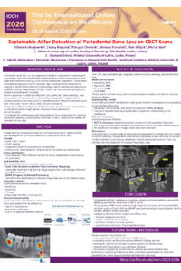
Periodontal bone loss is a key indicator of periodontitis severity and progression. Reliable detection is essential for timely intervention, yet manual assessment of cone-beam computed tomography (CBCT) scans is time-consuming and prone to interobserver variability. Artificial intelligence (AI), particularly deep learning, has shown promise in automating image interpretation; however, the “black-box” nature of many AI systems limits clinical trust. Explainable AI (XAI) techniques may enhance transparency by providing visual and quantitative insights into model reasoning. This study evaluates an XAI-enabled framework for automated detection of periodontal bone loss on CBCT scans and examines clinicians’ perceptions of its usability and interpretability.
Methodology:
A convolutional neural network (CNN) was trained on 2,500 annotated CBCT scans from 1,000 patients, labeled by two expert periodontists. XAI tools, including Gradient-weighted Class Activation Mapping (Grad-CAM) and SHapley Additive exPlanations (SHAP), were applied to highlight image regions contributing to model predictions. Model performance was assessed using sensitivity, specificity, F1-score, and AUC. To evaluate interpretability, 32 clinicians (18 periodontists, 14 radiologists) reviewed 120 AI-assisted cases. Perceptions were measured using a validated 5-point Likert-scale questionnaire adapted from the System Usability Scale and the Trust in Automation framework, complemented by two open-ended questions. Responses were analyzed using descriptive statistics and thematic coding.
Results:
The CNN achieved 91% sensitivity, 88% specificity, an F1-score of 0.895, and an AUC of 0.94. Grad-CAM and SHAP visualizations aligned with expert annotations in 87% of cases. Clinicians reported improved diagnostic confidence (mean 4.3/5) and perceived transparency (4.2/5), while 78% indicated that XAI outputs facilitated faster case review without increasing cognitive load.
Conclusions:
The proposed XAI-based system accurately detects periodontal bone loss on CBCT scans and provides interpretable visual feedback that supports clinician trust and decision-making. Future work will include prospective validation and workflow integration in routine periodontal diagnostics.
Keywords
Explainable AI
CBCT
Periodontal bone loss
Deep learning
Diagnostic accuracy
Poster
Oliwia%20Andrzejewska%20poster.pdf
Artificial Intelligence for Maxillary Sinus Pathology Detection on Cone–Beam CT: A Systematic Review of Expert-Consensus Studies
From Risk to Ready: A Five-Step Roadmap for Regulatory-Ready AI in Clinical Care