Event submissions
Published
This submission belongs to the session S2. Tumor Heterogeneity of the event The 5th International Electronic Conference on Cancers
Published date
05 Jun, 2026
Academic Editor
'%3e%3cpath%20d='M12.6657%2010.9104C12.4185%2010.325%2012.0599%209.79327%2011.6097%209.34482C11.1609%208.89508%2010.6293%208.5365%2010.0442%208.28888C10.0389%208.28626%2010.0337%208.28495%2010.0285%208.28233C10.8446%207.69279%2011.3752%206.73249%2011.3752%205.64904C11.3752%203.8542%209.92103%202.39999%208.1262%202.39999C6.33136%202.39999%204.87715%203.8542%204.87715%205.64904C4.87715%206.73249%205.40774%207.69279%206.22393%208.28364C6.21869%208.28626%206.21345%208.28757%206.20821%208.29019C5.62129%208.5378%205.09463%208.89284%204.64265%209.34613C4.1929%209.79493%203.83432%2010.3266%203.58671%2010.9117C3.34345%2011.4845%203.21226%2012.0987%203.20023%2012.7209C3.19988%2012.7349%203.20233%2012.7488%203.20744%2012.7619C3.21255%2012.7749%203.22022%2012.7867%203.22998%2012.7968C3.23975%2012.8068%203.25142%2012.8147%203.26431%2012.8202C3.2772%2012.8256%203.29105%2012.8284%203.30504%2012.8284H4.09109C4.14874%2012.8284%204.19459%2012.7825%204.1959%2012.7262C4.2221%2011.7148%204.62823%2010.7676%205.34617%2010.0497C6.08899%209.30683%207.0755%208.89808%208.1262%208.89808C9.17689%208.89808%2010.1634%209.30683%2010.9062%2010.0497C11.6242%2010.7676%2012.0303%2011.7148%2012.0565%2012.7262C12.0578%2012.7838%2012.1037%2012.8284%2012.1613%2012.8284H12.9474C12.9613%2012.8284%2012.9752%2012.8256%2012.9881%2012.8202C13.001%2012.8147%2013.0126%2012.8068%2013.0224%2012.7968C13.0322%2012.7867%2013.0398%2012.7749%2013.0449%2012.7619C13.0501%2012.7488%2013.0525%2012.7349%2013.0522%2012.7209C13.0391%2012.0947%2012.9094%2011.4855%2012.6657%2010.9104ZM8.1262%207.9024C7.52486%207.9024%206.9589%207.6679%206.53312%207.24211C6.10733%206.81633%205.87283%206.25037%205.87283%205.64904C5.87283%205.0477%206.10733%204.48174%206.53312%204.05596C6.9589%203.63018%207.52486%203.39567%208.1262%203.39567C8.72753%203.39567%209.29349%203.63018%209.71927%204.05596C10.1451%204.48174%2010.3796%205.0477%2010.3796%205.64904C10.3796%206.25037%2010.1451%206.81633%209.71927%207.24211C9.29349%207.6679%208.72753%207.9024%208.1262%207.9024Z'%20fill='%235D1EE1'/%3e%3c/g%3e%3c/svg%3e) Henry Heng
Henry HengCitation
Maria Karpova, Artur Bozhenko, Alexey Dolinin, Fedor Sarafanov, Nikolay Ilin, Evgeny Mareev, Irina Balalaeva, COMPARISON OF THE SENSITIVITY OF CANCER AND NORMAL HUMAN CELLS TO CHANGES IN BACKGROUND MAGNETIC FIELD, in Proceedings of The 5th International Electronic Conference on Cancers, 10 June–12 June 2026, MDPI: Basel, Switzerland
Share
Email
Facebook
Twitter
LinkedIn
COMPARISON OF THE SENSITIVITY OF CANCER AND NORMAL HUMAN CELLS TO CHANGES IN BACKGROUND MAGNETIC FIELD
'%3e%3cpath%20d='M12.6647%2010.9104C12.4176%2010.325%2012.0589%209.7933%2011.6088%209.34485C11.16%208.89511%2010.6283%208.53653%2010.0432%208.28891C10.038%208.28629%2010.0327%208.28498%2010.0275%208.28236C10.8437%207.69282%2011.3743%206.73252%2011.3743%205.64907C11.3743%203.85423%209.92005%202.40002%208.12522%202.40002C6.33038%202.40002%204.87618%203.85423%204.87618%205.64907C4.87618%206.73252%205.40677%207.69282%206.22296%208.28367C6.21772%208.28629%206.21248%208.2876%206.20724%208.29022C5.62031%208.53783%205.09365%208.89287%204.64167%209.34616C4.19192%209.79496%203.83334%2010.3266%203.58573%2010.9117C3.34247%2011.4846%203.21128%2012.0987%203.19925%2012.721C3.1989%2012.735%203.20135%2012.7489%203.20646%2012.7619C3.21157%2012.7749%203.21924%2012.7868%203.22901%2012.7968C3.23877%2012.8068%203.25045%2012.8148%203.26334%2012.8202C3.27623%2012.8256%203.29007%2012.8284%203.30406%2012.8284H4.09012C4.14776%2012.8284%204.19362%2012.7825%204.19493%2012.7262C4.22113%2011.7148%204.62726%2010.7676%205.34519%2010.0497C6.08802%209.30686%207.07452%208.89811%208.12522%208.89811C9.17592%208.89811%2010.1624%209.30686%2010.9052%2010.0497C11.6232%2010.7676%2012.0293%2011.7148%2012.0555%2012.7262C12.0568%2012.7839%2012.1027%2012.8284%2012.1603%2012.8284H12.9464C12.9604%2012.8284%2012.9742%2012.8256%2012.9871%2012.8202C13%2012.8148%2013.0117%2012.8068%2013.0214%2012.7968C13.0312%2012.7868%2013.0389%2012.7749%2013.044%2012.7619C13.0491%2012.7489%2013.0515%2012.735%2013.0512%2012.721C13.0381%2012.0947%2012.9084%2011.4856%2012.6647%2010.9104ZM8.12522%207.90243C7.52388%207.90243%206.95792%207.66793%206.53214%207.24215C6.10636%206.81636%205.87185%206.2504%205.87185%205.64907C5.87185%205.04773%206.10636%204.48177%206.53214%204.05599C6.95792%203.63021%207.52388%203.3957%208.12522%203.3957C8.72655%203.3957%209.29252%203.63021%209.7183%204.05599C10.1441%204.48177%2010.3786%205.04773%2010.3786%205.64907C10.3786%206.2504%2010.1441%206.81636%209.7183%207.24215C9.29252%207.66793%208.72655%207.90243%208.12522%207.90243Z'%20fill='%235D1EE1'/%3e%3c/g%3e%3c/svg%3e)
Maria Karpova 1
Artur Bozhenko 1
Alexey Dolinin 2
Fedor Sarafanov 2
Nikolay Ilin 2
Evgeny Mareev 2

Irina Balalaeva 1
1. Institute of Biology and Biomedicine, Lobachevsky State University, 23 Gagarin Ave., Nizhny Novgorod, 603022, Russia
2. Lobachevsky State University, 23 Gagarin Ave., Nizhny Novgorod, 603022, Russia
Abstract
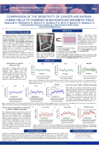
Weak magnetic fields (WMFs) with intensities up to 1 mT remain poorly studied, and the combined effects of magnetic fields (MFs) and other stress factors have received little attention.
The aim of this study was to investigate the effects of WMFs of different intensities on the growth of normal and cancer human cells, both as an independent factor and in combination with serum deprivation. The study used immortalized human embryonic kidney HEK-293T cells, cervical adenocarcinoma HeLa cells, and epidermoid carcinoma A-431 cells.
To generate the required magnetic conditions, a system of three-axis Helmholtz coils producing a gradient of a quasi-static MF with intensities from 6.5 to 115 μT was placed inside a CO₂ incubator. A 96-well plate containing the cells was positioned at the center of the setup.
Cells were exposed to the WMF for 72 hours, after which cell viability was assessed using the MTT assay. To evaluate the effect of serum deprivation on cellular sensitivity to WMFs, fetal bovine serum (FBS) concentration was reduced from 10% to 3% for HEK-293T cells and completely removed for HeLa and A-431 cells, resulting in a 50% decrease in viability in HEK-293T and HeLa cells and a 30% decrease in A-431 cells.
Proliferation of HEK-293T cells was dependent on MF intensity, and this effect was enhanced under stress conditions. No significant effects were observed in HeLa and A-431 cell lines, and this pattern persisted under serum deprivation. Tumor cells are known to exhibit pronounced metabolic and redox heterogeneity. It is suggested that MFs influence the cellular redox balance. The lack of effect in HeLa and A-431 may reflect the predominance of subpopulations in these lines that are adapted to oxidative stress.
This work was supported by a grant from Russian Science Foundation (RSF, project no. 25-24-00505, https://rscf.ru/project/25-24-00505/).
Keywords
magnetic field (MF)
weak magnetic field (WMF)
cell culture
cellular stress
nutritional deprivation
cancer cells.
Poster
Karpova et al_Poster.pdf
Synergistic Chemo-Immunomodulation via Nanocarrier Co-Delivery of Doxorubicin and Pexidartinib
Clinical outcomes of Sacituzumab Govitecan in metastatic breast cancer: evidence of survival and quality of life from a systematic review and meta analysis