Event submissions
Published
This submission belongs to the session S5. Causes, Diagnosis and Treatment of Cancer of the event The 5th International Electronic Conference on Cancers
Published date
05 Jun, 2026
Academic Editor
'%3e%3cpath%20d='M12.6657%2010.9104C12.4185%2010.325%2012.0599%209.79327%2011.6097%209.34482C11.1609%208.89508%2010.6293%208.5365%2010.0442%208.28888C10.0389%208.28626%2010.0337%208.28495%2010.0285%208.28233C10.8446%207.69279%2011.3752%206.73249%2011.3752%205.64904C11.3752%203.8542%209.92103%202.39999%208.1262%202.39999C6.33136%202.39999%204.87715%203.8542%204.87715%205.64904C4.87715%206.73249%205.40774%207.69279%206.22393%208.28364C6.21869%208.28626%206.21345%208.28757%206.20821%208.29019C5.62129%208.5378%205.09463%208.89284%204.64265%209.34613C4.1929%209.79493%203.83432%2010.3266%203.58671%2010.9117C3.34345%2011.4845%203.21226%2012.0987%203.20023%2012.7209C3.19988%2012.7349%203.20233%2012.7488%203.20744%2012.7619C3.21255%2012.7749%203.22022%2012.7867%203.22998%2012.7968C3.23975%2012.8068%203.25142%2012.8147%203.26431%2012.8202C3.2772%2012.8256%203.29105%2012.8284%203.30504%2012.8284H4.09109C4.14874%2012.8284%204.19459%2012.7825%204.1959%2012.7262C4.2221%2011.7148%204.62823%2010.7676%205.34617%2010.0497C6.08899%209.30683%207.0755%208.89808%208.1262%208.89808C9.17689%208.89808%2010.1634%209.30683%2010.9062%2010.0497C11.6242%2010.7676%2012.0303%2011.7148%2012.0565%2012.7262C12.0578%2012.7838%2012.1037%2012.8284%2012.1613%2012.8284H12.9474C12.9613%2012.8284%2012.9752%2012.8256%2012.9881%2012.8202C13.001%2012.8147%2013.0126%2012.8068%2013.0224%2012.7968C13.0322%2012.7867%2013.0398%2012.7749%2013.0449%2012.7619C13.0501%2012.7488%2013.0525%2012.7349%2013.0522%2012.7209C13.0391%2012.0947%2012.9094%2011.4855%2012.6657%2010.9104ZM8.1262%207.9024C7.52486%207.9024%206.9589%207.6679%206.53312%207.24211C6.10733%206.81633%205.87283%206.25037%205.87283%205.64904C5.87283%205.0477%206.10733%204.48174%206.53312%204.05596C6.9589%203.63018%207.52486%203.39567%208.1262%203.39567C8.72753%203.39567%209.29349%203.63018%209.71927%204.05596C10.1451%204.48174%2010.3796%205.0477%2010.3796%205.64904C10.3796%206.25037%2010.1451%206.81633%209.71927%207.24211C9.29349%207.6679%208.72753%207.9024%208.1262%207.9024Z'%20fill='%235D1EE1'/%3e%3c/g%3e%3c/svg%3e) Masaharu Seno
Masaharu SenoCitation
Mohammed El Magroud, mohammed meziane, Anass Haloui, Nassira Karich, Amal Bennani, SUBEPENDYMAL GIANT CELL ASTROCYTOMA: A RARE CENTRAL NERVOUS SYSTEM TUMOUR WITH A COMPLEX HISTOLOGY, in Proceedings of The 5th International Electronic Conference on Cancers, 10 June–12 June 2026, MDPI: Basel, Switzerland
Share
Email
Facebook
Twitter
LinkedIn
SUBEPENDYMAL GIANT CELL ASTROCYTOMA: A RARE CENTRAL NERVOUS SYSTEM TUMOUR WITH A COMPLEX HISTOLOGY
'%3e%3cpath%20d='M12.6647%2010.9104C12.4176%2010.325%2012.0589%209.7933%2011.6088%209.34485C11.16%208.89511%2010.6283%208.53653%2010.0432%208.28891C10.038%208.28629%2010.0327%208.28498%2010.0275%208.28236C10.8437%207.69282%2011.3743%206.73252%2011.3743%205.64907C11.3743%203.85423%209.92005%202.40002%208.12522%202.40002C6.33038%202.40002%204.87618%203.85423%204.87618%205.64907C4.87618%206.73252%205.40677%207.69282%206.22296%208.28367C6.21772%208.28629%206.21248%208.2876%206.20724%208.29022C5.62031%208.53783%205.09365%208.89287%204.64167%209.34616C4.19192%209.79496%203.83334%2010.3266%203.58573%2010.9117C3.34247%2011.4846%203.21128%2012.0987%203.19925%2012.721C3.1989%2012.735%203.20135%2012.7489%203.20646%2012.7619C3.21157%2012.7749%203.21924%2012.7868%203.22901%2012.7968C3.23877%2012.8068%203.25045%2012.8148%203.26334%2012.8202C3.27623%2012.8256%203.29007%2012.8284%203.30406%2012.8284H4.09012C4.14776%2012.8284%204.19362%2012.7825%204.19493%2012.7262C4.22113%2011.7148%204.62726%2010.7676%205.34519%2010.0497C6.08802%209.30686%207.07452%208.89811%208.12522%208.89811C9.17592%208.89811%2010.1624%209.30686%2010.9052%2010.0497C11.6232%2010.7676%2012.0293%2011.7148%2012.0555%2012.7262C12.0568%2012.7839%2012.1027%2012.8284%2012.1603%2012.8284H12.9464C12.9604%2012.8284%2012.9742%2012.8256%2012.9871%2012.8202C13%2012.8148%2013.0117%2012.8068%2013.0214%2012.7968C13.0312%2012.7868%2013.0389%2012.7749%2013.044%2012.7619C13.0491%2012.7489%2013.0515%2012.735%2013.0512%2012.721C13.0381%2012.0947%2012.9084%2011.4856%2012.6647%2010.9104ZM8.12522%207.90243C7.52388%207.90243%206.95792%207.66793%206.53214%207.24215C6.10636%206.81636%205.87185%206.2504%205.87185%205.64907C5.87185%205.04773%206.10636%204.48177%206.53214%204.05599C6.95792%203.63021%207.52388%203.3957%208.12522%203.3957C8.72655%203.3957%209.29252%203.63021%209.7183%204.05599C10.1441%204.48177%2010.3786%205.04773%2010.3786%205.64907C10.3786%206.2504%2010.1441%206.81636%209.7183%207.24215C9.29252%207.66793%208.72655%207.90243%208.12522%207.90243Z'%20fill='%235D1EE1'/%3e%3c/g%3e%3c/svg%3e)
Mohammed El Magroud 1
mohammed meziane 1
Anass Haloui 1
Nassira Karich 1
Amal Bennani 1
1. Anatomical Pathology Department, University Hospital Mohammed VI, Faculty of Medicine and Pharmacy, Oujda’s Mohammed Premier University, Oujda, Morocco
Abstract
Introduction
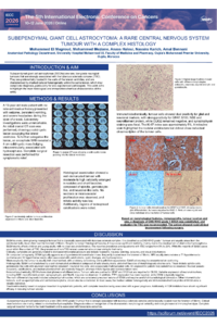
Subependymal giant cell astrocytomas (SEGAs) are rare, low-grade neuroglial tumours that are strongly associated with the tuberous sclerosis complex (TSC). They are preferentially located in the walls of the lateral ventricles, and are characterised by marked cellular heterogeneity within the same lesion, which may complicate diagnosis, particularly for less experienced pathologists. This work aims to highlight the main histological and immunohistochemical characteristics of this entity.
Methods and Results
A 13-year-old male patient with no relevant medical history presented with seizures, persistent vomiting, and severe headaches during the span of a week. Laboratory investigations were unremarkable, and an encephalic MRI revealed a 4 cm solid-cystic intraventricular mass associated with hydrocephalus. Complete surgical resection was performed for symptomatic relief.
Histological examination showed a well-demarcated tumour with moderate to high cellularity arranged in nodules and short fascicles, composed of spindle, gemistocyte-like, and neuronal-like cells. No necrosis or microvascular proliferation was observed, and mitotic activity was low.
Immunohistochemically, tumour cells showed dual positivity for glial and neuronal markers, with strong positivity for GFAP, S100, NSE and neurofilament protein, while OLIG2 remained negative, and synaptophysin staining was focal. The Ki-67 index was approximately 5%.
Based on morphological features, immunoprofile, and tumour location and size, a diagnosis of CNS WHO grade 1 SEGA was established, and evaluation for TSC was recommended. The patient showed rapid clinical improvement following surgery.
Conclusion
SEGA is a rare, slow-growing CNS WHO grade 1 tumour with symptoms that are mainly due to obstructive hydrocephalus. Diagnosis relies on recognition of its characteristic cellular morphological heterogeneity and dual glioneuronal differentiation. Complete surgical resection remains the cornerstone of treatment and is associated with favourable outcomes.
Keywords
subependymal giant cell astrocytoma
tuberous sclerosis complex
morphological heterogeneity
glioneuronal differentiation
Poster
SEGA IECC POSTER.pdf
Retrospective Registry for Uveal Melanoma in Lebanon: 10-year Single-Center Experience of a Major Referral Center in the Middle East and North Africa
Iron Oxide Nanoparticles as Potential Agents for Combined Radiotherapy