Event submissions
Published
This submission belongs to the session S2. Tumor Heterogeneity of the event The 5th International Electronic Conference on Cancers
Published date
05 Jun, 2026
Academic Editor
'%3e%3cpath%20d='M12.6657%2010.9104C12.4185%2010.325%2012.0599%209.79327%2011.6097%209.34482C11.1609%208.89508%2010.6293%208.5365%2010.0442%208.28888C10.0389%208.28626%2010.0337%208.28495%2010.0285%208.28233C10.8446%207.69279%2011.3752%206.73249%2011.3752%205.64904C11.3752%203.8542%209.92103%202.39999%208.1262%202.39999C6.33136%202.39999%204.87715%203.8542%204.87715%205.64904C4.87715%206.73249%205.40774%207.69279%206.22393%208.28364C6.21869%208.28626%206.21345%208.28757%206.20821%208.29019C5.62129%208.5378%205.09463%208.89284%204.64265%209.34613C4.1929%209.79493%203.83432%2010.3266%203.58671%2010.9117C3.34345%2011.4845%203.21226%2012.0987%203.20023%2012.7209C3.19988%2012.7349%203.20233%2012.7488%203.20744%2012.7619C3.21255%2012.7749%203.22022%2012.7867%203.22998%2012.7968C3.23975%2012.8068%203.25142%2012.8147%203.26431%2012.8202C3.2772%2012.8256%203.29105%2012.8284%203.30504%2012.8284H4.09109C4.14874%2012.8284%204.19459%2012.7825%204.1959%2012.7262C4.2221%2011.7148%204.62823%2010.7676%205.34617%2010.0497C6.08899%209.30683%207.0755%208.89808%208.1262%208.89808C9.17689%208.89808%2010.1634%209.30683%2010.9062%2010.0497C11.6242%2010.7676%2012.0303%2011.7148%2012.0565%2012.7262C12.0578%2012.7838%2012.1037%2012.8284%2012.1613%2012.8284H12.9474C12.9613%2012.8284%2012.9752%2012.8256%2012.9881%2012.8202C13.001%2012.8147%2013.0126%2012.8068%2013.0224%2012.7968C13.0322%2012.7867%2013.0398%2012.7749%2013.0449%2012.7619C13.0501%2012.7488%2013.0525%2012.7349%2013.0522%2012.7209C13.0391%2012.0947%2012.9094%2011.4855%2012.6657%2010.9104ZM8.1262%207.9024C7.52486%207.9024%206.9589%207.6679%206.53312%207.24211C6.10733%206.81633%205.87283%206.25037%205.87283%205.64904C5.87283%205.0477%206.10733%204.48174%206.53312%204.05596C6.9589%203.63018%207.52486%203.39567%208.1262%203.39567C8.72753%203.39567%209.29349%203.63018%209.71927%204.05596C10.1451%204.48174%2010.3796%205.0477%2010.3796%205.64904C10.3796%206.25037%2010.1451%206.81633%209.71927%207.24211C9.29349%207.6679%208.72753%207.9024%208.1262%207.9024Z'%20fill='%235D1EE1'/%3e%3c/g%3e%3c/svg%3e) Masaharu Seno
Masaharu SenoCitation
Manal KHARKHACH, asmae AISSAOUI, Anass HALOUI, Nassira KARICH, Amal BENNANI, Endometrioid Adenocarcinoma of the Endometrium with Mucinous Differentiation: An Anatomopathological Case Report, in Proceedings of The 5th International Electronic Conference on Cancers, 10 June–12 June 2026, MDPI: Basel, Switzerland
Share
Email
Facebook
Twitter
LinkedIn
Endometrioid Adenocarcinoma of the Endometrium with Mucinous Differentiation: An Anatomopathological Case Report
'%3e%3cpath%20d='M12.6647%2010.9104C12.4176%2010.325%2012.0589%209.7933%2011.6088%209.34485C11.16%208.89511%2010.6283%208.53653%2010.0432%208.28891C10.038%208.28629%2010.0327%208.28498%2010.0275%208.28236C10.8437%207.69282%2011.3743%206.73252%2011.3743%205.64907C11.3743%203.85423%209.92005%202.40002%208.12522%202.40002C6.33038%202.40002%204.87618%203.85423%204.87618%205.64907C4.87618%206.73252%205.40677%207.69282%206.22296%208.28367C6.21772%208.28629%206.21248%208.2876%206.20724%208.29022C5.62031%208.53783%205.09365%208.89287%204.64167%209.34616C4.19192%209.79496%203.83334%2010.3266%203.58573%2010.9117C3.34247%2011.4846%203.21128%2012.0987%203.19925%2012.721C3.1989%2012.735%203.20135%2012.7489%203.20646%2012.7619C3.21157%2012.7749%203.21924%2012.7868%203.22901%2012.7968C3.23877%2012.8068%203.25045%2012.8148%203.26334%2012.8202C3.27623%2012.8256%203.29007%2012.8284%203.30406%2012.8284H4.09012C4.14776%2012.8284%204.19362%2012.7825%204.19493%2012.7262C4.22113%2011.7148%204.62726%2010.7676%205.34519%2010.0497C6.08802%209.30686%207.07452%208.89811%208.12522%208.89811C9.17592%208.89811%2010.1624%209.30686%2010.9052%2010.0497C11.6232%2010.7676%2012.0293%2011.7148%2012.0555%2012.7262C12.0568%2012.7839%2012.1027%2012.8284%2012.1603%2012.8284H12.9464C12.9604%2012.8284%2012.9742%2012.8256%2012.9871%2012.8202C13%2012.8148%2013.0117%2012.8068%2013.0214%2012.7968C13.0312%2012.7868%2013.0389%2012.7749%2013.044%2012.7619C13.0491%2012.7489%2013.0515%2012.735%2013.0512%2012.721C13.0381%2012.0947%2012.9084%2011.4856%2012.6647%2010.9104ZM8.12522%207.90243C7.52388%207.90243%206.95792%207.66793%206.53214%207.24215C6.10636%206.81636%205.87185%206.2504%205.87185%205.64907C5.87185%205.04773%206.10636%204.48177%206.53214%204.05599C6.95792%203.63021%207.52388%203.3957%208.12522%203.3957C8.72655%203.3957%209.29252%203.63021%209.7183%204.05599C10.1441%204.48177%2010.3786%205.04773%2010.3786%205.64907C10.3786%206.2504%2010.1441%206.81636%209.7183%207.24215C9.29252%207.66793%208.72655%207.90243%208.12522%207.90243Z'%20fill='%235D1EE1'/%3e%3c/g%3e%3c/svg%3e)
Manal KHARKHACH 1
asmae AISSAOUI 1
Anass HALOUI 1
Nassira KARICH 1
Amal BENNANI 1
1. Pathology departement, Univercity hospital Mohammed VI, faculty of medecine and pharmacy, Mohamed 1 univversity, Oujda 60000, Morocco
Abstract
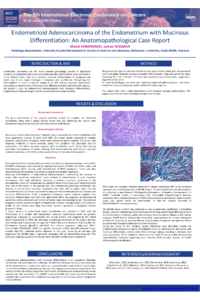
Endometrioid adenocarcinoma is the most common histological subtype of endometrial carcinoma. The presence of mucinous differentiation may represent a diagnostic challenge, particularly in distinguishing it from primary mucinous carcinoma of the endometrium or mixed epithelial tumors, entities that may carry different prognostic and therapeutic implications.
We report the case of a 63-year-old woman who underwent total hysterectomy with bilateral salpingo-oophorectomy for endometrial carcinoma. Histopathological examination revealed a FIGO grade 2 endometrioid adenocarcinoma with deep myometrial invasion exceeding 50% of the myometrial thickness. The tumor displayed predominantly glandular and papillary architecture, with tumor cells showing abundant cytoplasm containing intracytoplasmic mucin. Mucinous differentiation was confirmed by Alcian blue staining, which highlighted both intracytoplasmic and luminal mucin deposits. No serous or clear cell component was identified.
Immunohistochemical analysis demonstrated a wild-type p53 staining pattern and a deficient mismatch repair (dMMR) phenotype, characterized by loss of PMS2 expression with weak and heterogeneous MLH1 staining, while MSH2 and MSH6 were retained. The tumor cells showed diffuse positivity for CK7 and progesterone receptors, supporting an endometrial origin and excluding a high-grade serous carcinoma.
This case highlights the morphological and immunohistochemical features of endometrioid adenocarcinoma with mucinous differentiation and emphasizes the importance of an integrated diagnostic approach. Accurate recognition of this variant is essential to avoid misclassification and to ensure appropriate prognostic stratification and therapeutic management in accordance with current FIGO and ESGO/ESTRO/ESP recommendations.
Keywords
Endometrium
endometrioid adenocarcinoma
mucinous differentiation
immunohistochemistry
pathology
Poster
poster IECC KHARKHACH Manal.pdf
Extracellular vesicles as a targeted drug delivery platform for cancer treatment
Progression Control With Neoadjuvant DOS Versus SOX in HER2-Negative Locally Advanced Gastric Cancer: Multicenter Real-World Survival and Subgroup Evidence