Event submissions
Published
This submission belongs to the session S7. Novel Methods and Technologies for Research and Treatment of the event The 5th International Electronic Conference on Cancers
Published date
05 Jun, 2026
Academic Editor
'%3e%3cpath%20d='M12.6657%2010.9104C12.4185%2010.325%2012.0599%209.79327%2011.6097%209.34482C11.1609%208.89508%2010.6293%208.5365%2010.0442%208.28888C10.0389%208.28626%2010.0337%208.28495%2010.0285%208.28233C10.8446%207.69279%2011.3752%206.73249%2011.3752%205.64904C11.3752%203.8542%209.92103%202.39999%208.1262%202.39999C6.33136%202.39999%204.87715%203.8542%204.87715%205.64904C4.87715%206.73249%205.40774%207.69279%206.22393%208.28364C6.21869%208.28626%206.21345%208.28757%206.20821%208.29019C5.62129%208.5378%205.09463%208.89284%204.64265%209.34613C4.1929%209.79493%203.83432%2010.3266%203.58671%2010.9117C3.34345%2011.4845%203.21226%2012.0987%203.20023%2012.7209C3.19988%2012.7349%203.20233%2012.7488%203.20744%2012.7619C3.21255%2012.7749%203.22022%2012.7867%203.22998%2012.7968C3.23975%2012.8068%203.25142%2012.8147%203.26431%2012.8202C3.2772%2012.8256%203.29105%2012.8284%203.30504%2012.8284H4.09109C4.14874%2012.8284%204.19459%2012.7825%204.1959%2012.7262C4.2221%2011.7148%204.62823%2010.7676%205.34617%2010.0497C6.08899%209.30683%207.0755%208.89808%208.1262%208.89808C9.17689%208.89808%2010.1634%209.30683%2010.9062%2010.0497C11.6242%2010.7676%2012.0303%2011.7148%2012.0565%2012.7262C12.0578%2012.7838%2012.1037%2012.8284%2012.1613%2012.8284H12.9474C12.9613%2012.8284%2012.9752%2012.8256%2012.9881%2012.8202C13.001%2012.8147%2013.0126%2012.8068%2013.0224%2012.7968C13.0322%2012.7867%2013.0398%2012.7749%2013.0449%2012.7619C13.0501%2012.7488%2013.0525%2012.7349%2013.0522%2012.7209C13.0391%2012.0947%2012.9094%2011.4855%2012.6657%2010.9104ZM8.1262%207.9024C7.52486%207.9024%206.9589%207.6679%206.53312%207.24211C6.10733%206.81633%205.87283%206.25037%205.87283%205.64904C5.87283%205.0477%206.10733%204.48174%206.53312%204.05596C6.9589%203.63018%207.52486%203.39567%208.1262%203.39567C8.72753%203.39567%209.29349%203.63018%209.71927%204.05596C10.1451%204.48174%2010.3796%205.0477%2010.3796%205.64904C10.3796%206.25037%2010.1451%206.81633%209.71927%207.24211C9.29349%207.6679%208.72753%207.9024%208.1262%207.9024Z'%20fill='%235D1EE1'/%3e%3c/g%3e%3c/svg%3e) Guo-Min Li
Guo-Min LiCitation
Anastasia Dubrovina, Margarita Pustovalova, Sergey Leonov, Yulia Merkher, Development Omnipose Neural Network-Based Analytical Module for Morphometric Analysis of Metastatic Cancer Cells, in Proceedings of The 5th International Electronic Conference on Cancers, 10 June–12 June 2026, MDPI: Basel, Switzerland
Share
Email
Facebook
Twitter
LinkedIn
Development Omnipose Neural Network-Based Analytical Module for Morphometric Analysis of Metastatic Cancer Cells
'%3e%3cpath%20d='M12.6647%2010.9104C12.4176%2010.325%2012.0589%209.7933%2011.6088%209.34485C11.16%208.89511%2010.6283%208.53653%2010.0432%208.28891C10.038%208.28629%2010.0327%208.28498%2010.0275%208.28236C10.8437%207.69282%2011.3743%206.73252%2011.3743%205.64907C11.3743%203.85423%209.92005%202.40002%208.12522%202.40002C6.33038%202.40002%204.87618%203.85423%204.87618%205.64907C4.87618%206.73252%205.40677%207.69282%206.22296%208.28367C6.21772%208.28629%206.21248%208.2876%206.20724%208.29022C5.62031%208.53783%205.09365%208.89287%204.64167%209.34616C4.19192%209.79496%203.83334%2010.3266%203.58573%2010.9117C3.34247%2011.4846%203.21128%2012.0987%203.19925%2012.721C3.1989%2012.735%203.20135%2012.7489%203.20646%2012.7619C3.21157%2012.7749%203.21924%2012.7868%203.22901%2012.7968C3.23877%2012.8068%203.25045%2012.8148%203.26334%2012.8202C3.27623%2012.8256%203.29007%2012.8284%203.30406%2012.8284H4.09012C4.14776%2012.8284%204.19362%2012.7825%204.19493%2012.7262C4.22113%2011.7148%204.62726%2010.7676%205.34519%2010.0497C6.08802%209.30686%207.07452%208.89811%208.12522%208.89811C9.17592%208.89811%2010.1624%209.30686%2010.9052%2010.0497C11.6232%2010.7676%2012.0293%2011.7148%2012.0555%2012.7262C12.0568%2012.7839%2012.1027%2012.8284%2012.1603%2012.8284H12.9464C12.9604%2012.8284%2012.9742%2012.8256%2012.9871%2012.8202C13%2012.8148%2013.0117%2012.8068%2013.0214%2012.7968C13.0312%2012.7868%2013.0389%2012.7749%2013.044%2012.7619C13.0491%2012.7489%2013.0515%2012.735%2013.0512%2012.721C13.0381%2012.0947%2012.9084%2011.4856%2012.6647%2010.9104ZM8.12522%207.90243C7.52388%207.90243%206.95792%207.66793%206.53214%207.24215C6.10636%206.81636%205.87185%206.2504%205.87185%205.64907C5.87185%205.04773%206.10636%204.48177%206.53214%204.05599C6.95792%203.63021%207.52388%203.3957%208.12522%203.3957C8.72655%203.3957%209.29252%203.63021%209.7183%204.05599C10.1441%204.48177%2010.3786%205.04773%2010.3786%205.64907C10.3786%206.2504%2010.1441%206.81636%209.7183%207.24215C9.29252%207.66793%208.72655%207.90243%208.12522%207.90243Z'%20fill='%235D1EE1'/%3e%3c/g%3e%3c/svg%3e)
Anastasia Dubrovina 1

Margarita Pustovalova 1
Sergey Leonov 1

Yulia Merkher 1,2
1. Institute of Future Biophysics, Moscow Center for Basic Research, Moscow 123592, Russia
2. Department of Biomedical Engineering, Technion -- Israel Institute of Technology, Haifa 3200003, Israel
Abstract
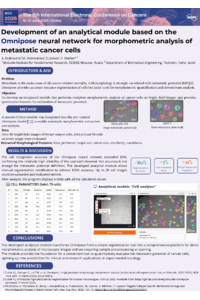
Introduction. Assessing metastatic potential (MP) based on morphological parameters of cancer cells is a promising direction in experimental oncology. The shape of a cell often correlates with an invasive phenotype, characterized by greater elongation and decreased roundness. A deep neural network image-segmentation algorithm implemented in Omnipose lacks tools for obtaining morphological data and their subsequent analysis for MP evaluation. Our aim was to develop a specialized software module for complete morphometric analysis of cells in microscopic images without need in specific pre-processing or staining.
Materials and Methods. Custom Python code (used packages: matplotlib.pyplot, skimage, scipy, google.colab, cellpose) was integrated into a pre-trained Omnipose model. The created module performs end-to-end analytics and calculation of key morphologic parameters: Area, Perimeter, Major_axis, Minor_axis, Circularity, Aspect_Ratio, Roundness, Solidity. The main innovation is an algorithm that analyzes these parameters, assigns characteristics to each cell, and outputs averaged values for further assessment of MP. Segmentation performance was evaluated by comparison with manual analysis using microscopic images of 65 breast cancer cells with high (MDA-MB-231) and low (MCF-7) MP.
Results. The Omnipose neural network's cell recognition accuracy exceeded 80%, confirming the high reliability of the approach. Measured morphologic parameters: Area, Perimeter, Major_axis, Minor_axis demonstrated strong agreement with manual analysis, with errors below 5%. The shape descriptors: Circularity, Solidity, Roundness and Aspect_Ratio demonstrated high concordance, with a maximum deviation of 11% from manually calculated values.
Conclusions. This work developed an analytical module that transforms Omnipose from a segmentation tool into a platform for direct morphometric analysis of microscopic images. The module serves as a foundation for a convenient tool to quantitatively evaluate cancer cell metastasis.
Keywords
Automated morphometric analysis
metastatic potential
image segmentation
breast cancer cells
cancer morphology biomarkers
Poster
170867 Poster (final).pdf
Time-Dependent SHAP Interpretation of a Multicenter ML DFS Model After Neoadjuvant Chemotherapy in Gastric Cancer
A Cross-Sectional Evaluation of Guideline-Adherent Screening Eligibility Among Zimbabwean Patients Diagnosed with Lung Cancer