Event submissions
Published
This submission belongs to the session S2. Bone Biomaterials of the event The 2nd International Online Conference on Functional Biomaterials
Published date
03 Jul, 2026
Academic Editor
'%3e%3cpath%20d='M12.6657%2010.9104C12.4185%2010.325%2012.0599%209.79327%2011.6097%209.34482C11.1609%208.89508%2010.6293%208.5365%2010.0442%208.28888C10.0389%208.28626%2010.0337%208.28495%2010.0285%208.28233C10.8446%207.69279%2011.3752%206.73249%2011.3752%205.64904C11.3752%203.8542%209.92103%202.39999%208.1262%202.39999C6.33136%202.39999%204.87715%203.8542%204.87715%205.64904C4.87715%206.73249%205.40774%207.69279%206.22393%208.28364C6.21869%208.28626%206.21345%208.28757%206.20821%208.29019C5.62129%208.5378%205.09463%208.89284%204.64265%209.34613C4.1929%209.79493%203.83432%2010.3266%203.58671%2010.9117C3.34345%2011.4845%203.21226%2012.0987%203.20023%2012.7209C3.19988%2012.7349%203.20233%2012.7488%203.20744%2012.7619C3.21255%2012.7749%203.22022%2012.7867%203.22998%2012.7968C3.23975%2012.8068%203.25142%2012.8147%203.26431%2012.8202C3.2772%2012.8256%203.29105%2012.8284%203.30504%2012.8284H4.09109C4.14874%2012.8284%204.19459%2012.7825%204.1959%2012.7262C4.2221%2011.7148%204.62823%2010.7676%205.34617%2010.0497C6.08899%209.30683%207.0755%208.89808%208.1262%208.89808C9.17689%208.89808%2010.1634%209.30683%2010.9062%2010.0497C11.6242%2010.7676%2012.0303%2011.7148%2012.0565%2012.7262C12.0578%2012.7838%2012.1037%2012.8284%2012.1613%2012.8284H12.9474C12.9613%2012.8284%2012.9752%2012.8256%2012.9881%2012.8202C13.001%2012.8147%2013.0126%2012.8068%2013.0224%2012.7968C13.0322%2012.7867%2013.0398%2012.7749%2013.0449%2012.7619C13.0501%2012.7488%2013.0525%2012.7349%2013.0522%2012.7209C13.0391%2012.0947%2012.9094%2011.4855%2012.6657%2010.9104ZM8.1262%207.9024C7.52486%207.9024%206.9589%207.6679%206.53312%207.24211C6.10733%206.81633%205.87283%206.25037%205.87283%205.64904C5.87283%205.0477%206.10733%204.48174%206.53312%204.05596C6.9589%203.63018%207.52486%203.39567%208.1262%203.39567C8.72753%203.39567%209.29349%203.63018%209.71927%204.05596C10.1451%204.48174%2010.3796%205.0477%2010.3796%205.64904C10.3796%206.25037%2010.1451%206.81633%209.71927%207.24211C9.29349%207.6679%208.72753%207.9024%208.1262%207.9024Z'%20fill='%235D1EE1'/%3e%3c/g%3e%3c/svg%3e) Elisa Boanini
Elisa BoaniniCitation
Smriti Aryal AC, Shahanas Kabeer, Md Sofiqul Islam, BMP7-Modified Polycaprolactone Nanofibrous Scaffolds Improve Osteoblast Activity, in Proceedings of The 2nd International Online Conference on Functional Biomaterials, 8 July–10 July 2026, MDPI: Basel, Switzerland
Share
Email
Facebook
Twitter
LinkedIn
BMP7-Modified Polycaprolactone Nanofibrous Scaffolds Improve Osteoblast Activity
'%3e%3cpath%20d='M12.6647%2010.9104C12.4176%2010.325%2012.0589%209.7933%2011.6088%209.34485C11.16%208.89511%2010.6283%208.53653%2010.0432%208.28891C10.038%208.28629%2010.0327%208.28498%2010.0275%208.28236C10.8437%207.69282%2011.3743%206.73252%2011.3743%205.64907C11.3743%203.85423%209.92005%202.40002%208.12522%202.40002C6.33038%202.40002%204.87618%203.85423%204.87618%205.64907C4.87618%206.73252%205.40677%207.69282%206.22296%208.28367C6.21772%208.28629%206.21248%208.2876%206.20724%208.29022C5.62031%208.53783%205.09365%208.89287%204.64167%209.34616C4.19192%209.79496%203.83334%2010.3266%203.58573%2010.9117C3.34247%2011.4846%203.21128%2012.0987%203.19925%2012.721C3.1989%2012.735%203.20135%2012.7489%203.20646%2012.7619C3.21157%2012.7749%203.21924%2012.7868%203.22901%2012.7968C3.23877%2012.8068%203.25045%2012.8148%203.26334%2012.8202C3.27623%2012.8256%203.29007%2012.8284%203.30406%2012.8284H4.09012C4.14776%2012.8284%204.19362%2012.7825%204.19493%2012.7262C4.22113%2011.7148%204.62726%2010.7676%205.34519%2010.0497C6.08802%209.30686%207.07452%208.89811%208.12522%208.89811C9.17592%208.89811%2010.1624%209.30686%2010.9052%2010.0497C11.6232%2010.7676%2012.0293%2011.7148%2012.0555%2012.7262C12.0568%2012.7839%2012.1027%2012.8284%2012.1603%2012.8284H12.9464C12.9604%2012.8284%2012.9742%2012.8256%2012.9871%2012.8202C13%2012.8148%2013.0117%2012.8068%2013.0214%2012.7968C13.0312%2012.7868%2013.0389%2012.7749%2013.044%2012.7619C13.0491%2012.7489%2013.0515%2012.735%2013.0512%2012.721C13.0381%2012.0947%2012.9084%2011.4856%2012.6647%2010.9104ZM8.12522%207.90243C7.52388%207.90243%206.95792%207.66793%206.53214%207.24215C6.10636%206.81636%205.87185%206.2504%205.87185%205.64907C5.87185%205.04773%206.10636%204.48177%206.53214%204.05599C6.95792%203.63021%207.52388%203.3957%208.12522%203.3957C8.72655%203.3957%209.29252%203.63021%209.7183%204.05599C10.1441%204.48177%2010.3786%205.04773%2010.3786%205.64907C10.3786%206.2504%2010.1441%206.81636%209.7183%207.24215C9.29252%207.66793%208.72655%207.90243%208.12522%207.90243Z'%20fill='%235D1EE1'/%3e%3c/g%3e%3c/svg%3e)
Smriti Aryal AC 1
Shahanas Kabeer 2

Md Sofiqul Islam 3
1. Department of Oral and Craniofacial Health Science, College of Dental Medicine, University of Sharjah, Sharjah, United Arab Emirates
2. Research Institute for Medical and Health Sciences, University of Sharjah, Sharjah, United Arab Emirates
3. Department of Operative Dentistry, RAK College of Dental Sciences, RAK Medical and Health Sciences University, Ras Al-Khaimah, 30248, United Arab Emirates
Abstract
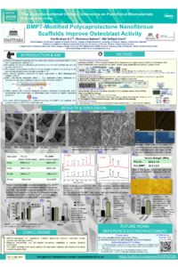
Introduction: Effective biomaterial integration with host bone cells remains a significant problem in the field of regenerative medicine. Biomaterials that act as both biological stimulants and structural scaffolds are required for bone healing. The polymer polycaprolactone (PCL) is strong enough to maintain its structure on its own, but lacks inherent osteo-inductive properties. One potential bioactive component for bone regeneration is Bone Morphogenetic Protein-7 (BMP7), which regulates osteoblast activity. This study aimed to create BMP7-integrated PCL scaffolds (PCL-BMP7) and evaluate their biological effects on MC3T3-E1 osteoblast cells.
Methods: PCL-BMP7 were fabricated via electrospinning with pure PCL membranes, whereas Pure PCL (PCL-Ct) was used as a control. Scaffold morphology and mechanical properties were characterized using scanning electron microscopy and tensile testing. Surface hydrophilicity was determined using contact angle analysis; molecular interactions were evaluated using X-ray diffraction and Raman spectroscopy. MC3T3-E1 cells were cultured on PCL-BMP7 and PCL-Ct. Cell attachment was observed by microscopy; MTT assay (“3- (4,5-dimethylthiazol-2-yl)-2,5-diphenyltetrazolium bromide assay”) was used to assess osteoblast viability. Wound healing, ALP (alkaline phosphatase activity) and mineral deposition were also evaluated by wound healing assay, ALP assay and alizarin red staining respectively. Data was analyzed by ANOVA and post hoc tests.
Results: In PCL-BMP7, SEM revealed a uniform fiber morphology and homogeneous BMP7 distribution. Raman and XRD analyses indicated enhanced molecular interactions. Contact angle measurements showed significantly improved hydrophilicity (**p<0.01). MC3T3-E1 cells exhibited excellent attachment and spreading on PCL-BMP7 scaffolds. Increased osteoblast cell viability was observed by the MTT assay at 24, 48, and 72 hours respectively (*p<0.05). Additionally, osteogenic capability and osteoblast wound healing were improved compared to Ct.
Conclusions: BMP7-functionalized PCL nanofibrous scaffolds improved osteoblast viability, healing capacity and osteogenic activity. These findings support the potential application of PCL-BMP7 scaffolds in regenerative dentistry and medicine.
Keywords
BMP7
PCL-BMP7
Osteoblast
Regenerative dentistry
medicine
Poster
Poster bmp7.pdf
Development and biological in vitro study of three-dimensional printed poly(lactic acid) scaffolds coated with gelatin-based coaxial nanofibrous coatings for bone repair
MXENE-MODIFIED TI/HAP COMPOSITES AS A BIOMATERIAL PLATFORM FOR BONE TISSUE REGENERATION