Event submissions
Published
This submission belongs to the session S4. Biomaterials for Tissue Engineering and Regenerative Medicine of the event The 2nd International Online Conference on Functional Biomaterials
Published date
03 Jul, 2026
Academic Editor
'%3e%3cpath%20d='M12.6657%2010.9104C12.4185%2010.325%2012.0599%209.79327%2011.6097%209.34482C11.1609%208.89508%2010.6293%208.5365%2010.0442%208.28888C10.0389%208.28626%2010.0337%208.28495%2010.0285%208.28233C10.8446%207.69279%2011.3752%206.73249%2011.3752%205.64904C11.3752%203.8542%209.92103%202.39999%208.1262%202.39999C6.33136%202.39999%204.87715%203.8542%204.87715%205.64904C4.87715%206.73249%205.40774%207.69279%206.22393%208.28364C6.21869%208.28626%206.21345%208.28757%206.20821%208.29019C5.62129%208.5378%205.09463%208.89284%204.64265%209.34613C4.1929%209.79493%203.83432%2010.3266%203.58671%2010.9117C3.34345%2011.4845%203.21226%2012.0987%203.20023%2012.7209C3.19988%2012.7349%203.20233%2012.7488%203.20744%2012.7619C3.21255%2012.7749%203.22022%2012.7867%203.22998%2012.7968C3.23975%2012.8068%203.25142%2012.8147%203.26431%2012.8202C3.2772%2012.8256%203.29105%2012.8284%203.30504%2012.8284H4.09109C4.14874%2012.8284%204.19459%2012.7825%204.1959%2012.7262C4.2221%2011.7148%204.62823%2010.7676%205.34617%2010.0497C6.08899%209.30683%207.0755%208.89808%208.1262%208.89808C9.17689%208.89808%2010.1634%209.30683%2010.9062%2010.0497C11.6242%2010.7676%2012.0303%2011.7148%2012.0565%2012.7262C12.0578%2012.7838%2012.1037%2012.8284%2012.1613%2012.8284H12.9474C12.9613%2012.8284%2012.9752%2012.8256%2012.9881%2012.8202C13.001%2012.8147%2013.0126%2012.8068%2013.0224%2012.7968C13.0322%2012.7867%2013.0398%2012.7749%2013.0449%2012.7619C13.0501%2012.7488%2013.0525%2012.7349%2013.0522%2012.7209C13.0391%2012.0947%2012.9094%2011.4855%2012.6657%2010.9104ZM8.1262%207.9024C7.52486%207.9024%206.9589%207.6679%206.53312%207.24211C6.10733%206.81633%205.87283%206.25037%205.87283%205.64904C5.87283%205.0477%206.10733%204.48174%206.53312%204.05596C6.9589%203.63018%207.52486%203.39567%208.1262%203.39567C8.72753%203.39567%209.29349%203.63018%209.71927%204.05596C10.1451%204.48174%2010.3796%205.0477%2010.3796%205.64904C10.3796%206.25037%2010.1451%206.81633%209.71927%207.24211C9.29349%207.6679%208.72753%207.9024%208.1262%207.9024Z'%20fill='%235D1EE1'/%3e%3c/g%3e%3c/svg%3e) Piergiorgio Gentile
Piergiorgio GentileCitation
Ayşenur Atak, Ayşen Güngör, YUKSEL CETİN, Ayça Bal Öztürk, İlke Gürol, Banu Mansuroğlu, Evaluation of a Biomimetic and Electroactive 3D Bioprinted Cardiac Patch: From Bioink Optimization to Functional Regeneration in a Perfusion System, in Proceedings of The 2nd International Online Conference on Functional Biomaterials, 8 July–10 July 2026, MDPI: Basel, Switzerland
Share
Email
Facebook
Twitter
LinkedIn
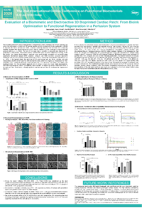
Evaluation of a Biomimetic and Electroactive 3D Bioprinted Cardiac Patch: From Bioink Optimization to Functional Regeneration in a Perfusion System
'%3e%3cpath%20d='M12.6647%2010.9104C12.4176%2010.325%2012.0589%209.7933%2011.6088%209.34485C11.16%208.89511%2010.6283%208.53653%2010.0432%208.28891C10.038%208.28629%2010.0327%208.28498%2010.0275%208.28236C10.8437%207.69282%2011.3743%206.73252%2011.3743%205.64907C11.3743%203.85423%209.92005%202.40002%208.12522%202.40002C6.33038%202.40002%204.87618%203.85423%204.87618%205.64907C4.87618%206.73252%205.40677%207.69282%206.22296%208.28367C6.21772%208.28629%206.21248%208.2876%206.20724%208.29022C5.62031%208.53783%205.09365%208.89287%204.64167%209.34616C4.19192%209.79496%203.83334%2010.3266%203.58573%2010.9117C3.34247%2011.4846%203.21128%2012.0987%203.19925%2012.721C3.1989%2012.735%203.20135%2012.7489%203.20646%2012.7619C3.21157%2012.7749%203.21924%2012.7868%203.22901%2012.7968C3.23877%2012.8068%203.25045%2012.8148%203.26334%2012.8202C3.27623%2012.8256%203.29007%2012.8284%203.30406%2012.8284H4.09012C4.14776%2012.8284%204.19362%2012.7825%204.19493%2012.7262C4.22113%2011.7148%204.62726%2010.7676%205.34519%2010.0497C6.08802%209.30686%207.07452%208.89811%208.12522%208.89811C9.17592%208.89811%2010.1624%209.30686%2010.9052%2010.0497C11.6232%2010.7676%2012.0293%2011.7148%2012.0555%2012.7262C12.0568%2012.7839%2012.1027%2012.8284%2012.1603%2012.8284H12.9464C12.9604%2012.8284%2012.9742%2012.8256%2012.9871%2012.8202C13%2012.8148%2013.0117%2012.8068%2013.0214%2012.7968C13.0312%2012.7868%2013.0389%2012.7749%2013.044%2012.7619C13.0491%2012.7489%2013.0515%2012.735%2013.0512%2012.721C13.0381%2012.0947%2012.9084%2011.4856%2012.6647%2010.9104ZM8.12522%207.90243C7.52388%207.90243%206.95792%207.66793%206.53214%207.24215C6.10636%206.81636%205.87185%206.2504%205.87185%205.64907C5.87185%205.04773%206.10636%204.48177%206.53214%204.05599C6.95792%203.63021%207.52388%203.3957%208.12522%203.3957C8.72655%203.3957%209.29252%203.63021%209.7183%204.05599C10.1441%204.48177%2010.3786%205.04773%2010.3786%205.64907C10.3786%206.2504%2010.1441%206.81636%209.7183%207.24215C9.29252%207.66793%208.72655%207.90243%208.12522%207.90243Z'%20fill='%235D1EE1'/%3e%3c/g%3e%3c/svg%3e)
Ayşenur Atak 1,2
Ayşen Güngör 2,3
YUKSEL CETİN 4
Ayça Bal Öztürk 3,5,6
İlke Gürol 2
Banu Mansuroğlu 1
1. Molecular Biology and Genetics, Yildiz Technical University, Istanbul, Türkiye
2. Life Sciences, Biotechnology R&D, TUBITAK MAM, Gebze, Kocaeli, Türkiye
3. Department of Stem Cell and Tissue Engineering, Institute of Health Sciences, Istinye University, Istanbul, Türkiye
4. The Scientific and Technological Research Council (TUBITAK), Marmara Research Center (MAM), Gebze, Kocaeli, Türkiye
5. Stem Cell and Tissue Engineering Application and Research Center (ISUKOK), Istinye University, Istanbul, Türkiye
6. Department of Analytical Chemistry, Faculty of Pharmacy, Istinye University, Istanbul, Türkiye
Abstract
The escalating demand for functional myocardial tissue following ischemic injury has catalyzed the development of biomimetic and electroactive 3D cardiac patches. In this study, a hybrid bioink—comprising decellularized bovine pericardium (dECM), silk fibroin (SF), alginate, and gold nanoparticles (AuNPs)—was developed and systematically optimized for extrusion-based 3D bioprinting to fabricate structurally robust and electrophysiologically active constructs. The bioink was engineered to recapitulate the biochemical complexity of the native extracellular matrix (ECM) while providing essential mechanical reinforcement and conductive properties to facilitate synchronized cardiac tissue regeneration. In the subsequent phase, the biodegradability, biocompatibility, and regenerative efficacy of the 3D-bioprinted patches were evaluated within a dynamic 3D perfusion culture system. Under physiologically relevant flow conditions, the constructs were assessed for their capacity to support cellular adhesion, proliferation, and hierarchical tissue organization. The functional role of the patches in cardiac reconstruction was evaluated through a multi-parametric approach, including biochemical assays, histological assessments, and immunofluorescence analyses of cardiac-specific biomarkers and ECM remodelling. Furthermore, the safety profile of the construct was rigorously established through ISO-standardized cell viability assays, in vitro irritation/corrosion tests, and in vitro skin sensitization assessments. The degradation kinetics and structural integrity were systematically monitored to ensure controlled biodegradability. Our findings demonstrate that this hybrid 3D cardiac patch offers a biomimetic microenvironment with favorable mechanical–electroactive properties and high biosafety, supporting cardiac maturation within a perfusion environment. This study provides a comprehensive framework for the development of multifunctional cardiac constructs and underscores their translational potential in regenerative cardiovascular medicine.
Keywords
3D bioprinting
Alginate
dECM
Silk fibroin
NanoAu
Cardiac Tissue Engineering
Poster
sciforum Poster-175379.pdf
From Spontaneous Differentiation to Functional Synaptic Networks: Neurogenic Modulation of SH‑SY5Y Cells by Semiconductive P3HT Interfaces
Hybrid 3D-Printed/Electrospun Honeycomb PCL Scaffolds Improve Mechanical Stability and Promote Osteoblast Colonization