Event submissions
Published
This submission belongs to the session S2. Surveillance and Epidemiological Modelling at the Human–Animal–Environment Interface of the event The 2nd International Online Conference on Veterinary Sciences
Published date
02 Sep, 2026
Academic Editor
'%3e%3cpath%20d='M12.6657%2010.9104C12.4185%2010.325%2012.0599%209.79327%2011.6097%209.34482C11.1609%208.89508%2010.6293%208.5365%2010.0442%208.28888C10.0389%208.28626%2010.0337%208.28495%2010.0285%208.28233C10.8446%207.69279%2011.3752%206.73249%2011.3752%205.64904C11.3752%203.8542%209.92103%202.39999%208.1262%202.39999C6.33136%202.39999%204.87715%203.8542%204.87715%205.64904C4.87715%206.73249%205.40774%207.69279%206.22393%208.28364C6.21869%208.28626%206.21345%208.28757%206.20821%208.29019C5.62129%208.5378%205.09463%208.89284%204.64265%209.34613C4.1929%209.79493%203.83432%2010.3266%203.58671%2010.9117C3.34345%2011.4845%203.21226%2012.0987%203.20023%2012.7209C3.19988%2012.7349%203.20233%2012.7488%203.20744%2012.7619C3.21255%2012.7749%203.22022%2012.7867%203.22998%2012.7968C3.23975%2012.8068%203.25142%2012.8147%203.26431%2012.8202C3.2772%2012.8256%203.29105%2012.8284%203.30504%2012.8284H4.09109C4.14874%2012.8284%204.19459%2012.7825%204.1959%2012.7262C4.2221%2011.7148%204.62823%2010.7676%205.34617%2010.0497C6.08899%209.30683%207.0755%208.89808%208.1262%208.89808C9.17689%208.89808%2010.1634%209.30683%2010.9062%2010.0497C11.6242%2010.7676%2012.0303%2011.7148%2012.0565%2012.7262C12.0578%2012.7838%2012.1037%2012.8284%2012.1613%2012.8284H12.9474C12.9613%2012.8284%2012.9752%2012.8256%2012.9881%2012.8202C13.001%2012.8147%2013.0126%2012.8068%2013.0224%2012.7968C13.0322%2012.7867%2013.0398%2012.7749%2013.0449%2012.7619C13.0501%2012.7488%2013.0525%2012.7349%2013.0522%2012.7209C13.0391%2012.0947%2012.9094%2011.4855%2012.6657%2010.9104ZM8.1262%207.9024C7.52486%207.9024%206.9589%207.6679%206.53312%207.24211C6.10733%206.81633%205.87283%206.25037%205.87283%205.64904C5.87283%205.0477%206.10733%204.48174%206.53312%204.05596C6.9589%203.63018%207.52486%203.39567%208.1262%203.39567C8.72753%203.39567%209.29349%203.63018%209.71927%204.05596C10.1451%204.48174%2010.3796%205.0477%2010.3796%205.64904C10.3796%206.25037%2010.1451%206.81633%209.71927%207.24211C9.29349%207.6679%208.72753%207.9024%208.1262%207.9024Z'%20fill='%235D1EE1'/%3e%3c/g%3e%3c/svg%3e) Francesco Mira
Francesco MiraCitation
Zahra Sadat Yaghoubi Kolouri, Shahabeddin Safi, Splenic and Systemic Lymphoma in an FIV‑Positive Cat: A Case Report Highlighting Diagnostic Discordance in Cytology Interpretation, in Proceedings of The 2nd International Online Conference on Veterinary Sciences, 7 September–9 September 2026, MDPI: Basel, Switzerland
Share
Email
Facebook
Twitter
LinkedIn
Splenic and Systemic Lymphoma in an FIV‑Positive Cat: A Case Report Highlighting Diagnostic Discordance in Cytology Interpretation
'%3e%3cpath%20d='M12.6647%2010.9104C12.4176%2010.325%2012.0589%209.7933%2011.6088%209.34485C11.16%208.89511%2010.6283%208.53653%2010.0432%208.28891C10.038%208.28629%2010.0327%208.28498%2010.0275%208.28236C10.8437%207.69282%2011.3743%206.73252%2011.3743%205.64907C11.3743%203.85423%209.92005%202.40002%208.12522%202.40002C6.33038%202.40002%204.87618%203.85423%204.87618%205.64907C4.87618%206.73252%205.40677%207.69282%206.22296%208.28367C6.21772%208.28629%206.21248%208.2876%206.20724%208.29022C5.62031%208.53783%205.09365%208.89287%204.64167%209.34616C4.19192%209.79496%203.83334%2010.3266%203.58573%2010.9117C3.34247%2011.4846%203.21128%2012.0987%203.19925%2012.721C3.1989%2012.735%203.20135%2012.7489%203.20646%2012.7619C3.21157%2012.7749%203.21924%2012.7868%203.22901%2012.7968C3.23877%2012.8068%203.25045%2012.8148%203.26334%2012.8202C3.27623%2012.8256%203.29007%2012.8284%203.30406%2012.8284H4.09012C4.14776%2012.8284%204.19362%2012.7825%204.19493%2012.7262C4.22113%2011.7148%204.62726%2010.7676%205.34519%2010.0497C6.08802%209.30686%207.07452%208.89811%208.12522%208.89811C9.17592%208.89811%2010.1624%209.30686%2010.9052%2010.0497C11.6232%2010.7676%2012.0293%2011.7148%2012.0555%2012.7262C12.0568%2012.7839%2012.1027%2012.8284%2012.1603%2012.8284H12.9464C12.9604%2012.8284%2012.9742%2012.8256%2012.9871%2012.8202C13%2012.8148%2013.0117%2012.8068%2013.0214%2012.7968C13.0312%2012.7868%2013.0389%2012.7749%2013.044%2012.7619C13.0491%2012.7489%2013.0515%2012.735%2013.0512%2012.721C13.0381%2012.0947%2012.9084%2011.4856%2012.6647%2010.9104ZM8.12522%207.90243C7.52388%207.90243%206.95792%207.66793%206.53214%207.24215C6.10636%206.81636%205.87185%206.2504%205.87185%205.64907C5.87185%205.04773%206.10636%204.48177%206.53214%204.05599C6.95792%203.63021%207.52388%203.3957%208.12522%203.3957C8.72655%203.3957%209.29252%203.63021%209.7183%204.05599C10.1441%204.48177%2010.3786%205.04773%2010.3786%205.64907C10.3786%206.2504%2010.1441%206.81636%209.7183%207.24215C9.29252%207.66793%208.72655%207.90243%208.12522%207.90243Z'%20fill='%235D1EE1'/%3e%3c/g%3e%3c/svg%3e)
Zahra Sadat Yaghoubi Kolouri 1
Shahabeddin Safi 1
1. Department of Clinical Pathology, Faculty of Specialized Veterinary Sciences, Science and Research Branch, Islamic Azad University, Tehran, Iran
Abstract
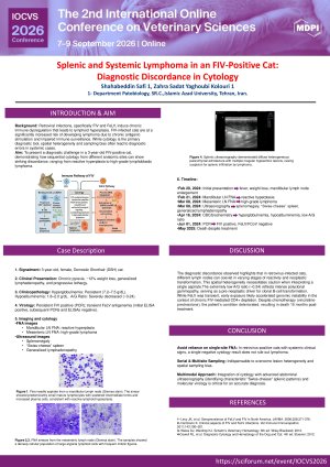
Feline immunodeficiency virus (FIV) causes chronic immune dysregulation that provokes lymphoid hyperplasia, often complicating the cytologic diagnosis of neoplasia. We present a 3-year-old female domestic shorthair cat from Iran evaluated for chronic pyrexia, approximately 12% weight loss, and generalized lymphadenopathy. Ultrasonography revealed marked splenomegaly with a “Swiss-cheese” or mottled parenchymal pattern, hepatomegaly, and bilateral renal medullary rim signs. Hematological analysis demonstrated persistent hyperglobulinemia, hypoalbuminemia, and a markedly decreased albumin-to-globulin (A/G) ratio (~0.24), indicative of sustained antigenic stimulation. Virologic testing confirmed persistent FIV infection via PCR. Notably, a single initial FeLV ELISA was positive, but subsequent PCRs were negative, suggesting transient antigenemia or false-positive detection. Cytologic evaluations of different lymph nodes yielded highly discordant results: mandibular aspirates indicated reactive lymphoid hyperplasia, whereas mesenteric aspirates 16 days later revealed high-grade lymphoblastic lymphoma. This striking disparity illustrates profound lesion heterogeneity and spatial sampling bias under retroviral immunosuppression. Despite partial initial responsiveness to a vincristine-prednisolone-based chemotherapy protocol and supportive care, the patient’s condition deteriorated, culminating in death 15 months post-diagnosis. This case emphasizes the diagnostic challenges of differentiating FIV-induced reactivity from lymphoma. It highlights the necessity of serial, multisite cytologic sampling integrated with advanced imaging and precise virologic testing for the accurate diagnosis of feline hematopoietic malignancies.
Keywords
FIV
FeLV
Lymphoma
Splenic lesions
Cytology.
Poster
FIV case report 6.Edited.pdf
Cracking the Shell: Genomic Insights into Antimicrobial-Resistant Escherichia coli on Retail Eggs in the United Arab Emirates
De Novo Discovery of Dual Hantavirus Gc/Gn Inhibitors Using a General-Purpose Generative AI