Event submissions
Published
This submission belongs to the session S1. One Health Approaches to Emerging Zoonotic Threats of the event The 2nd International Online Conference on Veterinary Sciences
Published date
02 Sep, 2026
Academic Editor
'%3e%3cpath%20d='M12.6657%2010.9104C12.4185%2010.325%2012.0599%209.79327%2011.6097%209.34482C11.1609%208.89508%2010.6293%208.5365%2010.0442%208.28888C10.0389%208.28626%2010.0337%208.28495%2010.0285%208.28233C10.8446%207.69279%2011.3752%206.73249%2011.3752%205.64904C11.3752%203.8542%209.92103%202.39999%208.1262%202.39999C6.33136%202.39999%204.87715%203.8542%204.87715%205.64904C4.87715%206.73249%205.40774%207.69279%206.22393%208.28364C6.21869%208.28626%206.21345%208.28757%206.20821%208.29019C5.62129%208.5378%205.09463%208.89284%204.64265%209.34613C4.1929%209.79493%203.83432%2010.3266%203.58671%2010.9117C3.34345%2011.4845%203.21226%2012.0987%203.20023%2012.7209C3.19988%2012.7349%203.20233%2012.7488%203.20744%2012.7619C3.21255%2012.7749%203.22022%2012.7867%203.22998%2012.7968C3.23975%2012.8068%203.25142%2012.8147%203.26431%2012.8202C3.2772%2012.8256%203.29105%2012.8284%203.30504%2012.8284H4.09109C4.14874%2012.8284%204.19459%2012.7825%204.1959%2012.7262C4.2221%2011.7148%204.62823%2010.7676%205.34617%2010.0497C6.08899%209.30683%207.0755%208.89808%208.1262%208.89808C9.17689%208.89808%2010.1634%209.30683%2010.9062%2010.0497C11.6242%2010.7676%2012.0303%2011.7148%2012.0565%2012.7262C12.0578%2012.7838%2012.1037%2012.8284%2012.1613%2012.8284H12.9474C12.9613%2012.8284%2012.9752%2012.8256%2012.9881%2012.8202C13.001%2012.8147%2013.0126%2012.8068%2013.0224%2012.7968C13.0322%2012.7867%2013.0398%2012.7749%2013.0449%2012.7619C13.0501%2012.7488%2013.0525%2012.7349%2013.0522%2012.7209C13.0391%2012.0947%2012.9094%2011.4855%2012.6657%2010.9104ZM8.1262%207.9024C7.52486%207.9024%206.9589%207.6679%206.53312%207.24211C6.10733%206.81633%205.87283%206.25037%205.87283%205.64904C5.87283%205.0477%206.10733%204.48174%206.53312%204.05596C6.9589%203.63018%207.52486%203.39567%208.1262%203.39567C8.72753%203.39567%209.29349%203.63018%209.71927%204.05596C10.1451%204.48174%2010.3796%205.0477%2010.3796%205.64904C10.3796%206.25037%2010.1451%206.81633%209.71927%207.24211C9.29349%207.6679%208.72753%207.9024%208.1262%207.9024Z'%20fill='%235D1EE1'/%3e%3c/g%3e%3c/svg%3e) Vittorio Sarchese
Vittorio SarcheseCitation
Maryam Kermanshahian, Tara Marzban, Artificial Intelligence for Chest Radiography in Tuberculosis Detection Across Humans and Animals: A Systematic Review of One Health Diagnostic Applications, in Proceedings of The 2nd International Online Conference on Veterinary Sciences, 7 September–9 September 2026, MDPI: Basel, Switzerland
Share
Email
Facebook
Twitter
LinkedIn
Artificial Intelligence for Chest Radiography in Tuberculosis Detection Across Humans and Animals: A Systematic Review of One Health Diagnostic Applications
'%3e%3cpath%20d='M12.6647%2010.9104C12.4176%2010.325%2012.0589%209.7933%2011.6088%209.34485C11.16%208.89511%2010.6283%208.53653%2010.0432%208.28891C10.038%208.28629%2010.0327%208.28498%2010.0275%208.28236C10.8437%207.69282%2011.3743%206.73252%2011.3743%205.64907C11.3743%203.85423%209.92005%202.40002%208.12522%202.40002C6.33038%202.40002%204.87618%203.85423%204.87618%205.64907C4.87618%206.73252%205.40677%207.69282%206.22296%208.28367C6.21772%208.28629%206.21248%208.2876%206.20724%208.29022C5.62031%208.53783%205.09365%208.89287%204.64167%209.34616C4.19192%209.79496%203.83334%2010.3266%203.58573%2010.9117C3.34247%2011.4846%203.21128%2012.0987%203.19925%2012.721C3.1989%2012.735%203.20135%2012.7489%203.20646%2012.7619C3.21157%2012.7749%203.21924%2012.7868%203.22901%2012.7968C3.23877%2012.8068%203.25045%2012.8148%203.26334%2012.8202C3.27623%2012.8256%203.29007%2012.8284%203.30406%2012.8284H4.09012C4.14776%2012.8284%204.19362%2012.7825%204.19493%2012.7262C4.22113%2011.7148%204.62726%2010.7676%205.34519%2010.0497C6.08802%209.30686%207.07452%208.89811%208.12522%208.89811C9.17592%208.89811%2010.1624%209.30686%2010.9052%2010.0497C11.6232%2010.7676%2012.0293%2011.7148%2012.0555%2012.7262C12.0568%2012.7839%2012.1027%2012.8284%2012.1603%2012.8284H12.9464C12.9604%2012.8284%2012.9742%2012.8256%2012.9871%2012.8202C13%2012.8148%2013.0117%2012.8068%2013.0214%2012.7968C13.0312%2012.7868%2013.0389%2012.7749%2013.044%2012.7619C13.0491%2012.7489%2013.0515%2012.735%2013.0512%2012.721C13.0381%2012.0947%2012.9084%2011.4856%2012.6647%2010.9104ZM8.12522%207.90243C7.52388%207.90243%206.95792%207.66793%206.53214%207.24215C6.10636%206.81636%205.87185%206.2504%205.87185%205.64907C5.87185%205.04773%206.10636%204.48177%206.53214%204.05599C6.95792%203.63021%207.52388%203.3957%208.12522%203.3957C8.72655%203.3957%209.29252%203.63021%209.7183%204.05599C10.1441%204.48177%2010.3786%205.04773%2010.3786%205.64907C10.3786%206.2504%2010.1441%206.81636%209.7183%207.24215C9.29252%207.66793%208.72655%207.90243%208.12522%207.90243Z'%20fill='%235D1EE1'/%3e%3c/g%3e%3c/svg%3e)
Maryam Kermanshahian 1
Tara Marzban 1
1. Department of Veterinary Medicine, School of Veterinary Medicine, Islamic Azad University, Babol Branch, Babol, Iran
Abstract
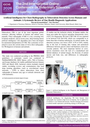
Background/Objective: Tuberculosis (TB) is one of the most important global zoonoses, affecting millions of human and animal cases annually. Chest radiography (CXR) is a key screening tool in human medicine, and in recent years, deep learning-based artificial intelligence (AI) algorithms have been developed for automated interpretation of CXR. This systematic review aimed to assess the current status of AI in chest radiography for TB diagnosis in humans and animals.
Methods: This study was conducted according to the PRISMA 2020 guidelines. A systematic search was conducted in PubMed/MEDLINE, IEEE Xplore, arXiv, Web of Science, and Scopus databases for studies published between January 2020 and March 2026. Inclusion criteria included original studies (excluding reviews and commentaries) that used deep learning algorithms (such as convolutional neural networks – CNN) to detect tuberculosis from chest radiographs in humans (any age) or animals (domestic and wild mammals).
Results: 23 studies met the inclusion criteria. In human studies, the mean area under the receiver operating characteristic curve (AUC) for diagnosing TB from CXR with AI was reported to be between 0.94 and 0.98, which was comparable to or better than that of human radiologists. In animal studies, the detection accuracy was lower (AUC: 0.76 to 0.85), mainly due to the lack of training data volume and anatomical differences between species (chest wall thickness, heart size, vascular pattern). The most important barriers to cross-species translation were) the lack of a standardized and labeled database of animal images, the diversity of animal species and body size, and differences in radiographic protocols.
Conclusion: Artificial intelligence in the diagnosis of TB from chest radiographs in humans has reached a high level of maturity, but its application in animals is very limited and nascent. Transfer learning from human to animal models represents a promising strategy to fill this gap.
Keywords
Artificial Intelligence
Chest X-ray
Deep Learning
One Health
Tuberculosis
Poster
Poster.pdf
Cow Nutrition as a Preventive Component of Antimicrobial Resistance Control and Food Safety in Cattle Production: A One Health Evidence Synthesis.
Development of an HR2-Derived Fusion Inhibitor Against Canine Coronavirus; in vitro and in silico approaches