Event submissions
Published
This submission belongs to the session 4. Molecular and Cellular Mechanisms, Comparative Toxicology, and Multi-Omics Integration of the event The 3rd International Online Conference on Toxics
Published date
04 Sep, 2026
Academic Editor
'%3e%3cpath%20d='M12.6657%2010.9104C12.4185%2010.325%2012.0599%209.79327%2011.6097%209.34482C11.1609%208.89508%2010.6293%208.5365%2010.0442%208.28888C10.0389%208.28626%2010.0337%208.28495%2010.0285%208.28233C10.8446%207.69279%2011.3752%206.73249%2011.3752%205.64904C11.3752%203.8542%209.92103%202.39999%208.1262%202.39999C6.33136%202.39999%204.87715%203.8542%204.87715%205.64904C4.87715%206.73249%205.40774%207.69279%206.22393%208.28364C6.21869%208.28626%206.21345%208.28757%206.20821%208.29019C5.62129%208.5378%205.09463%208.89284%204.64265%209.34613C4.1929%209.79493%203.83432%2010.3266%203.58671%2010.9117C3.34345%2011.4845%203.21226%2012.0987%203.20023%2012.7209C3.19988%2012.7349%203.20233%2012.7488%203.20744%2012.7619C3.21255%2012.7749%203.22022%2012.7867%203.22998%2012.7968C3.23975%2012.8068%203.25142%2012.8147%203.26431%2012.8202C3.2772%2012.8256%203.29105%2012.8284%203.30504%2012.8284H4.09109C4.14874%2012.8284%204.19459%2012.7825%204.1959%2012.7262C4.2221%2011.7148%204.62823%2010.7676%205.34617%2010.0497C6.08899%209.30683%207.0755%208.89808%208.1262%208.89808C9.17689%208.89808%2010.1634%209.30683%2010.9062%2010.0497C11.6242%2010.7676%2012.0303%2011.7148%2012.0565%2012.7262C12.0578%2012.7838%2012.1037%2012.8284%2012.1613%2012.8284H12.9474C12.9613%2012.8284%2012.9752%2012.8256%2012.9881%2012.8202C13.001%2012.8147%2013.0126%2012.8068%2013.0224%2012.7968C13.0322%2012.7867%2013.0398%2012.7749%2013.0449%2012.7619C13.0501%2012.7488%2013.0525%2012.7349%2013.0522%2012.7209C13.0391%2012.0947%2012.9094%2011.4855%2012.6657%2010.9104ZM8.1262%207.9024C7.52486%207.9024%206.9589%207.6679%206.53312%207.24211C6.10733%206.81633%205.87283%206.25037%205.87283%205.64904C5.87283%205.0477%206.10733%204.48174%206.53312%204.05596C6.9589%203.63018%207.52486%203.39567%208.1262%203.39567C8.72753%203.39567%209.29349%203.63018%209.71927%204.05596C10.1451%204.48174%2010.3796%205.0477%2010.3796%205.64904C10.3796%206.25037%2010.1451%206.81633%209.71927%207.24211C9.29349%207.6679%208.72753%207.9024%208.1262%207.9024Z'%20fill='%235D1EE1'/%3e%3c/g%3e%3c/svg%3e) Yankai Xia
Yankai XiaCitation
Madson Silveira de Melo, Maico Roberto Luckmann, Evelise Maria Nazari, Pyriproxyfen disrupts liver development in Gallus domesticus embryos at environmentally relevant concentrations, in Proceedings of The 3rd International Online Conference on Toxics, 9 September–11 September 2026, MDPI: Basel, Switzerland
Share
Email
Facebook
Twitter
LinkedIn
Pyriproxyfen disrupts liver development in Gallus domesticus embryos at environmentally relevant concentrations
'%3e%3cpath%20d='M12.6647%2010.9104C12.4176%2010.325%2012.0589%209.7933%2011.6088%209.34485C11.16%208.89511%2010.6283%208.53653%2010.0432%208.28891C10.038%208.28629%2010.0327%208.28498%2010.0275%208.28236C10.8437%207.69282%2011.3743%206.73252%2011.3743%205.64907C11.3743%203.85423%209.92005%202.40002%208.12522%202.40002C6.33038%202.40002%204.87618%203.85423%204.87618%205.64907C4.87618%206.73252%205.40677%207.69282%206.22296%208.28367C6.21772%208.28629%206.21248%208.2876%206.20724%208.29022C5.62031%208.53783%205.09365%208.89287%204.64167%209.34616C4.19192%209.79496%203.83334%2010.3266%203.58573%2010.9117C3.34247%2011.4846%203.21128%2012.0987%203.19925%2012.721C3.1989%2012.735%203.20135%2012.7489%203.20646%2012.7619C3.21157%2012.7749%203.21924%2012.7868%203.22901%2012.7968C3.23877%2012.8068%203.25045%2012.8148%203.26334%2012.8202C3.27623%2012.8256%203.29007%2012.8284%203.30406%2012.8284H4.09012C4.14776%2012.8284%204.19362%2012.7825%204.19493%2012.7262C4.22113%2011.7148%204.62726%2010.7676%205.34519%2010.0497C6.08802%209.30686%207.07452%208.89811%208.12522%208.89811C9.17592%208.89811%2010.1624%209.30686%2010.9052%2010.0497C11.6232%2010.7676%2012.0293%2011.7148%2012.0555%2012.7262C12.0568%2012.7839%2012.1027%2012.8284%2012.1603%2012.8284H12.9464C12.9604%2012.8284%2012.9742%2012.8256%2012.9871%2012.8202C13%2012.8148%2013.0117%2012.8068%2013.0214%2012.7968C13.0312%2012.7868%2013.0389%2012.7749%2013.044%2012.7619C13.0491%2012.7489%2013.0515%2012.735%2013.0512%2012.721C13.0381%2012.0947%2012.9084%2011.4856%2012.6647%2010.9104ZM8.12522%207.90243C7.52388%207.90243%206.95792%207.66793%206.53214%207.24215C6.10636%206.81636%205.87185%206.2504%205.87185%205.64907C5.87185%205.04773%206.10636%204.48177%206.53214%204.05599C6.95792%203.63021%207.52388%203.3957%208.12522%203.3957C8.72655%203.3957%209.29252%203.63021%209.7183%204.05599C10.1441%204.48177%2010.3786%205.04773%2010.3786%205.64907C10.3786%206.2504%2010.1441%206.81636%209.7183%207.24215C9.29252%207.66793%208.72655%207.90243%208.12522%207.90243Z'%20fill='%235D1EE1'/%3e%3c/g%3e%3c/svg%3e)
Madson Silveira de Melo 1
Maico Roberto Luckmann 1
Evelise Maria Nazari 1
1. Department of Cell Biology, Embryology and Genetics, Federal University of Santa Catarina, Florianópolis, Santa Catarina 88040-900, Brazil
Abstract
Introduction
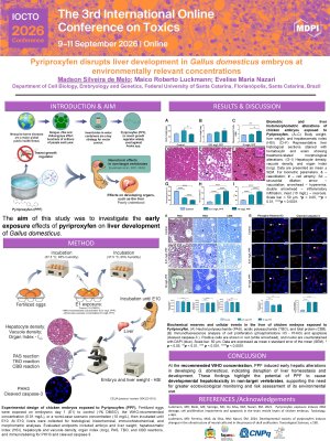
Extensively used in vector-control programs across public health and agriculture, pyriproxyfen (PPF) is an insect growth regulator considered to pose minimal risk to non-target organisms. However, growing evidence indicates that it can cross biological barriers and interfere with conserved molecular pathways. Although its neurotoxicity in vertebrates has been described, their hepatotoxic potential during embryogenesis remains poorly understood. Thus, the aim of this study was to investigate the PPF effects on liver development of Gallus domesticus.
Methods
Fertilized eggs were exposed at embryonic day 1 (E1) to vehicle control (1% DMSO), the WHO-recommended PPF concentration (0.01 mg/L), or a worst-case scenario concentration (10 mg/L). Embryos were incubated until E10, when liver were collected for biometric, histological, histochemical, and cellular analyses. All experimental procedures were approved by the Animal Use Ethics Committee of the Federal University of Santa Catarina (5843231018).
Results/Discussion
Embryos exposed to PPF exhibited increased hepatosomatic index along with marked histoarchitectural disruption, including reduced hepatocyte density and increased vacuolization at both PPF concentrations. The combined histopathological index was significantly elevated in PPF-exposed groups. Histochemical analyses further demonstrated metabolic impairment, whereas toluidine blue O reaction showed increased metachromasia at both PPF concentrations, suggesting accumulation of acidic components associated with cellular stress. Conversely, Periodic Acid–Schiff reaction revealed reduced neutral polysaccharides only at 10 mg/L, suggesting glycogen depletion. Reduced Coomassie Brilliant Blue reaction suggested loss of protein integrity or synthesis. Moreover, cellular markers confirmed disrupted hepatic development, in which proliferation (PHH3) was significantly reduced, while apoptosis (cleaved caspase-3) increased even at PPF environmentally relevant concentration (0.01 mg/L).
Conclusion
Together, these results demonstrate that PPF compromises liver homeostasis, metabolism, and cellular turnover during early development, establishing its potential as an embryonic hepatotoxicant. These findings underscore the need for ecotoxicological monitoring and stricter evaluation of PPF use in environments shared with non-target vertebrates.
Keywords
Embryotoxicity
Developmental toxicity
Larvicide totoxicity
Hepatotoxicity
Citotoxicology
Poster
Melo et al. 2026_IOCT.pdf
Zinc modulates nickel-induced oxidative stress in the liver of murine model
Di-n-butyl Phthalate-Induced Cardiovascular Effects Following Long-Term In Vitro Exposure