Event submissions
Published
This submission belongs to the session 4. Molecular and Cellular Mechanisms, Comparative Toxicology, and Multi-Omics Integration of the event The 3rd International Online Conference on Toxics
Published date
04 Sep, 2026
Academic Editor
'%3e%3cpath%20d='M12.6657%2010.9104C12.4185%2010.325%2012.0599%209.79327%2011.6097%209.34482C11.1609%208.89508%2010.6293%208.5365%2010.0442%208.28888C10.0389%208.28626%2010.0337%208.28495%2010.0285%208.28233C10.8446%207.69279%2011.3752%206.73249%2011.3752%205.64904C11.3752%203.8542%209.92103%202.39999%208.1262%202.39999C6.33136%202.39999%204.87715%203.8542%204.87715%205.64904C4.87715%206.73249%205.40774%207.69279%206.22393%208.28364C6.21869%208.28626%206.21345%208.28757%206.20821%208.29019C5.62129%208.5378%205.09463%208.89284%204.64265%209.34613C4.1929%209.79493%203.83432%2010.3266%203.58671%2010.9117C3.34345%2011.4845%203.21226%2012.0987%203.20023%2012.7209C3.19988%2012.7349%203.20233%2012.7488%203.20744%2012.7619C3.21255%2012.7749%203.22022%2012.7867%203.22998%2012.7968C3.23975%2012.8068%203.25142%2012.8147%203.26431%2012.8202C3.2772%2012.8256%203.29105%2012.8284%203.30504%2012.8284H4.09109C4.14874%2012.8284%204.19459%2012.7825%204.1959%2012.7262C4.2221%2011.7148%204.62823%2010.7676%205.34617%2010.0497C6.08899%209.30683%207.0755%208.89808%208.1262%208.89808C9.17689%208.89808%2010.1634%209.30683%2010.9062%2010.0497C11.6242%2010.7676%2012.0303%2011.7148%2012.0565%2012.7262C12.0578%2012.7838%2012.1037%2012.8284%2012.1613%2012.8284H12.9474C12.9613%2012.8284%2012.9752%2012.8256%2012.9881%2012.8202C13.001%2012.8147%2013.0126%2012.8068%2013.0224%2012.7968C13.0322%2012.7867%2013.0398%2012.7749%2013.0449%2012.7619C13.0501%2012.7488%2013.0525%2012.7349%2013.0522%2012.7209C13.0391%2012.0947%2012.9094%2011.4855%2012.6657%2010.9104ZM8.1262%207.9024C7.52486%207.9024%206.9589%207.6679%206.53312%207.24211C6.10733%206.81633%205.87283%206.25037%205.87283%205.64904C5.87283%205.0477%206.10733%204.48174%206.53312%204.05596C6.9589%203.63018%207.52486%203.39567%208.1262%203.39567C8.72753%203.39567%209.29349%203.63018%209.71927%204.05596C10.1451%204.48174%2010.3796%205.0477%2010.3796%205.64904C10.3796%206.25037%2010.1451%206.81633%209.71927%207.24211C9.29349%207.6679%208.72753%207.9024%208.1262%207.9024Z'%20fill='%235D1EE1'/%3e%3c/g%3e%3c/svg%3e) Yankai Xia
Yankai XiaCitation
Sini Macheri, Erica Bruce, Investigation of Cellular and Molecular Mechanisms of PFAS-Induced Lung Inflammation in a Human Alveolar Epithelial Cell Line, in Proceedings of The 3rd International Online Conference on Toxics, 9 September–11 September 2026, MDPI: Basel, Switzerland
Share
Email
Facebook
Twitter
LinkedIn
Investigation of Cellular and Molecular Mechanisms of PFAS-Induced Lung Inflammation in a Human Alveolar Epithelial Cell Line
'%3e%3cpath%20d='M12.6647%2010.9104C12.4176%2010.325%2012.0589%209.7933%2011.6088%209.34485C11.16%208.89511%2010.6283%208.53653%2010.0432%208.28891C10.038%208.28629%2010.0327%208.28498%2010.0275%208.28236C10.8437%207.69282%2011.3743%206.73252%2011.3743%205.64907C11.3743%203.85423%209.92005%202.40002%208.12522%202.40002C6.33038%202.40002%204.87618%203.85423%204.87618%205.64907C4.87618%206.73252%205.40677%207.69282%206.22296%208.28367C6.21772%208.28629%206.21248%208.2876%206.20724%208.29022C5.62031%208.53783%205.09365%208.89287%204.64167%209.34616C4.19192%209.79496%203.83334%2010.3266%203.58573%2010.9117C3.34247%2011.4846%203.21128%2012.0987%203.19925%2012.721C3.1989%2012.735%203.20135%2012.7489%203.20646%2012.7619C3.21157%2012.7749%203.21924%2012.7868%203.22901%2012.7968C3.23877%2012.8068%203.25045%2012.8148%203.26334%2012.8202C3.27623%2012.8256%203.29007%2012.8284%203.30406%2012.8284H4.09012C4.14776%2012.8284%204.19362%2012.7825%204.19493%2012.7262C4.22113%2011.7148%204.62726%2010.7676%205.34519%2010.0497C6.08802%209.30686%207.07452%208.89811%208.12522%208.89811C9.17592%208.89811%2010.1624%209.30686%2010.9052%2010.0497C11.6232%2010.7676%2012.0293%2011.7148%2012.0555%2012.7262C12.0568%2012.7839%2012.1027%2012.8284%2012.1603%2012.8284H12.9464C12.9604%2012.8284%2012.9742%2012.8256%2012.9871%2012.8202C13%2012.8148%2013.0117%2012.8068%2013.0214%2012.7968C13.0312%2012.7868%2013.0389%2012.7749%2013.044%2012.7619C13.0491%2012.7489%2013.0515%2012.735%2013.0512%2012.721C13.0381%2012.0947%2012.9084%2011.4856%2012.6647%2010.9104ZM8.12522%207.90243C7.52388%207.90243%206.95792%207.66793%206.53214%207.24215C6.10636%206.81636%205.87185%206.2504%205.87185%205.64907C5.87185%205.04773%206.10636%204.48177%206.53214%204.05599C6.95792%203.63021%207.52388%203.3957%208.12522%203.3957C8.72655%203.3957%209.29252%203.63021%209.7183%204.05599C10.1441%204.48177%2010.3786%205.04773%2010.3786%205.64907C10.3786%206.2504%2010.1441%206.81636%209.7183%207.24215C9.29252%207.66793%208.72655%207.90243%208.12522%207.90243Z'%20fill='%235D1EE1'/%3e%3c/g%3e%3c/svg%3e)
Sini Macheri 1
Erica Bruce 1
1. Environmental Science, Baylor University, Waco, TX
Abstract
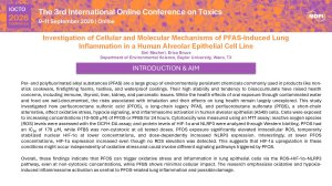
Per- and polyfluorinated alkyl substances (PFAS) are a large group of environmentally persistent chemicals widely used in products like non-stick cookware, firefighting foams, textiles, and waterproof coatings. Their high stability and tendency to bioaccumulate have raised health concerns, linked to immune, thyroid, liver, kidney, and pancreatic issues. Although the health effects of oral exposure through contaminated water and food are well-documented, the risks of inhalation and their impact on lung health remain largely unexplored. This study examined how perfluorooctane sulfonic acid (PFOS), a long-chain legacy PFAS, and perfluorobutane sulfonate (PFBS), a short-chain alternative, influence oxidative stress, hypoxia signaling, and inflammasome activation in human alveolar epithelial (A549) cells. Cells were exposed for 24 hours to increasing concentrations (10–200 µM) of PFOS or PFBS. Cytotoxicity was assessed using an MTT assay, reactive oxygen species (ROS) levels were measured with the DCFH-DA assay, and protein levels of HIF-1α and NLRP3 were analyzed via Western blotting and immunocytochemistry. PFOS had an IC₅₀ of 179 µM, while PFBS was non-cytotoxic at all tested doses. PFOS exposure significantly elevated intracellular ROS, caused a temporary nuclear stabilization of HIF-1α at lower concentrations, and dose-dependently increased NLRP3 expression—peaking at 100 µM and slightly decreasing at 150 µM. Immunocytochemistry showed increased cytoplasmic and perinuclear localization of NLRP3 following PFOS treatment. Overall, these results suggest that PFOS, even at non-cytotoxic levels, can induce oxidative stress and inflammatory pathways in lung epithelial cells through activation of the ROS–HIF-1α–NLRP3 axis, whereas PFBS exhibits minimal cellular effects. The study highlights oxidative and hypoxia-driven inflammasome activation as a key mechanism behind PFOS-related lung inflammation and potential injury.
Keywords
PFAS
invitro
inhalation
oxidative stress
cytotoxicity
Poster
SINI TOXICS SLIDES.pdf
THE PROTECTIVE EFFECTS OF N-ACETYLCYSTEINE ON DI-ETHYL PHTHALATE EXPOSED UTERUS OF WISTAR RATS
Biochemical Disruption of Glutathione-Dependent and Independent Antioxidant Defense Systems under Synergistic Pesticide Exposure in Trogoderma granarium