Event submissions
Published
This submission belongs to the session 4. Molecular and Cellular Mechanisms, Comparative Toxicology, and Multi-Omics Integration of the event The 3rd International Online Conference on Toxics
Published date
04 Sep, 2026
Academic Editor
'%3e%3cpath%20d='M12.6657%2010.9104C12.4185%2010.325%2012.0599%209.79327%2011.6097%209.34482C11.1609%208.89508%2010.6293%208.5365%2010.0442%208.28888C10.0389%208.28626%2010.0337%208.28495%2010.0285%208.28233C10.8446%207.69279%2011.3752%206.73249%2011.3752%205.64904C11.3752%203.8542%209.92103%202.39999%208.1262%202.39999C6.33136%202.39999%204.87715%203.8542%204.87715%205.64904C4.87715%206.73249%205.40774%207.69279%206.22393%208.28364C6.21869%208.28626%206.21345%208.28757%206.20821%208.29019C5.62129%208.5378%205.09463%208.89284%204.64265%209.34613C4.1929%209.79493%203.83432%2010.3266%203.58671%2010.9117C3.34345%2011.4845%203.21226%2012.0987%203.20023%2012.7209C3.19988%2012.7349%203.20233%2012.7488%203.20744%2012.7619C3.21255%2012.7749%203.22022%2012.7867%203.22998%2012.7968C3.23975%2012.8068%203.25142%2012.8147%203.26431%2012.8202C3.2772%2012.8256%203.29105%2012.8284%203.30504%2012.8284H4.09109C4.14874%2012.8284%204.19459%2012.7825%204.1959%2012.7262C4.2221%2011.7148%204.62823%2010.7676%205.34617%2010.0497C6.08899%209.30683%207.0755%208.89808%208.1262%208.89808C9.17689%208.89808%2010.1634%209.30683%2010.9062%2010.0497C11.6242%2010.7676%2012.0303%2011.7148%2012.0565%2012.7262C12.0578%2012.7838%2012.1037%2012.8284%2012.1613%2012.8284H12.9474C12.9613%2012.8284%2012.9752%2012.8256%2012.9881%2012.8202C13.001%2012.8147%2013.0126%2012.8068%2013.0224%2012.7968C13.0322%2012.7867%2013.0398%2012.7749%2013.0449%2012.7619C13.0501%2012.7488%2013.0525%2012.7349%2013.0522%2012.7209C13.0391%2012.0947%2012.9094%2011.4855%2012.6657%2010.9104ZM8.1262%207.9024C7.52486%207.9024%206.9589%207.6679%206.53312%207.24211C6.10733%206.81633%205.87283%206.25037%205.87283%205.64904C5.87283%205.0477%206.10733%204.48174%206.53312%204.05596C6.9589%203.63018%207.52486%203.39567%208.1262%203.39567C8.72753%203.39567%209.29349%203.63018%209.71927%204.05596C10.1451%204.48174%2010.3796%205.0477%2010.3796%205.64904C10.3796%206.25037%2010.1451%206.81633%209.71927%207.24211C9.29349%207.6679%208.72753%207.9024%208.1262%207.9024Z'%20fill='%235D1EE1'/%3e%3c/g%3e%3c/svg%3e) Yankai Xia
Yankai XiaCitation
Marco Aurelio M. Freire, Daniel Falcao, Walace Gomes-Leal, Rafael R. Lima, Chronic methylmercury exposure alters astrocytic morphology and NADPH diaphorase reactivity in the rat frontal cortex, in Proceedings of The 3rd International Online Conference on Toxics, 9 September–11 September 2026, MDPI: Basel, Switzerland
Share
Email
Facebook
Twitter
LinkedIn
Chronic methylmercury exposure alters astrocytic morphology and NADPH diaphorase reactivity in the rat frontal cortex
'%3e%3cpath%20d='M12.6647%2010.9104C12.4176%2010.325%2012.0589%209.7933%2011.6088%209.34485C11.16%208.89511%2010.6283%208.53653%2010.0432%208.28891C10.038%208.28629%2010.0327%208.28498%2010.0275%208.28236C10.8437%207.69282%2011.3743%206.73252%2011.3743%205.64907C11.3743%203.85423%209.92005%202.40002%208.12522%202.40002C6.33038%202.40002%204.87618%203.85423%204.87618%205.64907C4.87618%206.73252%205.40677%207.69282%206.22296%208.28367C6.21772%208.28629%206.21248%208.2876%206.20724%208.29022C5.62031%208.53783%205.09365%208.89287%204.64167%209.34616C4.19192%209.79496%203.83334%2010.3266%203.58573%2010.9117C3.34247%2011.4846%203.21128%2012.0987%203.19925%2012.721C3.1989%2012.735%203.20135%2012.7489%203.20646%2012.7619C3.21157%2012.7749%203.21924%2012.7868%203.22901%2012.7968C3.23877%2012.8068%203.25045%2012.8148%203.26334%2012.8202C3.27623%2012.8256%203.29007%2012.8284%203.30406%2012.8284H4.09012C4.14776%2012.8284%204.19362%2012.7825%204.19493%2012.7262C4.22113%2011.7148%204.62726%2010.7676%205.34519%2010.0497C6.08802%209.30686%207.07452%208.89811%208.12522%208.89811C9.17592%208.89811%2010.1624%209.30686%2010.9052%2010.0497C11.6232%2010.7676%2012.0293%2011.7148%2012.0555%2012.7262C12.0568%2012.7839%2012.1027%2012.8284%2012.1603%2012.8284H12.9464C12.9604%2012.8284%2012.9742%2012.8256%2012.9871%2012.8202C13%2012.8148%2013.0117%2012.8068%2013.0214%2012.7968C13.0312%2012.7868%2013.0389%2012.7749%2013.044%2012.7619C13.0491%2012.7489%2013.0515%2012.735%2013.0512%2012.721C13.0381%2012.0947%2012.9084%2011.4856%2012.6647%2010.9104ZM8.12522%207.90243C7.52388%207.90243%206.95792%207.66793%206.53214%207.24215C6.10636%206.81636%205.87185%206.2504%205.87185%205.64907C5.87185%205.04773%206.10636%204.48177%206.53214%204.05599C6.95792%203.63021%207.52388%203.3957%208.12522%203.3957C8.72655%203.3957%209.29252%203.63021%209.7183%204.05599C10.1441%204.48177%2010.3786%205.04773%2010.3786%205.64907C10.3786%206.2504%2010.1441%206.81636%209.7183%207.24215C9.29252%207.66793%208.72655%207.90243%208.12522%207.90243Z'%20fill='%235D1EE1'/%3e%3c/g%3e%3c/svg%3e)
Marco Aurelio M. Freire 1
Daniel Falcao 2
Walace Gomes-Leal 3

Rafael R. Lima 1
1. Laboratory of Functional and Structural Biology, Institute of Biological Sciences, Federal University of Pará, Belém 66075-110 PA, Brazil
2. VCU Health Systems, Virginia Commonwealth University, Richmond 23284 VA, USA
3. Laboratory of Experimental Neuroprotection and Neuroregeneration, Institute of Collective Health, Federal University of Western Pará, Santarém 68040-470 PA, Brazil
Abstract
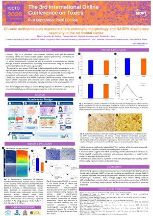
Introduction: Mercury (Hg) remains a persistent environmental pollutant with well-documented neurotoxic potential. Despite global awareness of its hazards, Hg continues to be extensively employed in artisanal gold mining, particularly in developing regions, contributing to environmental contamination and human exposure. In aquatic environments, inorganic Hg can be converted into methylmercury (MeHg) through microbial biomethylation, facilitating its bioaccumulation along the food chain and increasing the risk of human exposure. The central nervous system is particularly vulnerable to MeHg toxicity due to its high metabolic demand and sensitivity to oxidative and metabolic disturbances. Here, we assessed the effects of chronic MeHg exposure on NADPH-diaphorase (NADPH-d) activity and astrocytic responses in the frontal cortex of rats. Methods: Adult rats were randomly assigned to control and MeHg-exposed groups (n = 6 per group). The MeHg group received the compound by oral gavage (0.04 mg/kg/day) for 60 days, while control animals received the vehicle following the same schedule, in accordance with a protocol approved by the UFPA’s Ethics Committee for Animal Experimentation (ID# BIO 225-14-CEPAE-UFPA). After the exposure period, brain tissue samples were analyzed to determine Hg concentration, NADPH-d histochemical reactivity, and astrocytic morphology using glial fibrillary acidic protein (GFAP) immunohistochemistry. Results: Chronic MeHg exposure resulted in significant accumulation of Hg in cortical tissue and a reduction in NADPH-d neuropil optical density (p<0.05). Morphometric analysis indicated that NADPH-d-positive neurons were relatively preserved, suggesting resistance to chronic toxic exposure. In contrast, astrocytes exhibited marked morphological alterations, including cellular swelling and hypertrophy. Discussion/Conclusions: These findings suggest that prolonged exposure to MeHg induces selective neurochemical and glial changes in the frontal cortex. The reduction in NADPH-d neuropil reactivity may reflect impaired synthesis or intracellular transport of the enzyme, whereas the apparent resistance of NADPH-d-positive neurons could be related to metabolic adaptations or potential protective mechanisms involving nitric oxide signaling.
Keywords
Methylmercury
Neurotoxicity
Astrocytes
NADPH diaphorase
Frontal cortex
Poster
Freire et al. Poster IOCTO2026.pdf
Systems-level effects of plant-based cognitive enhancer extracts: A multi-omics perspective
An Inflamed Gut is Not Always a Leaky Gut: Mycotoxin Permeability in a Co-culture Model