Event submissions
Published
This submission belongs to the session 1. Exposure Routes / Exposome of Emerging Contaminants and Materials in the Environment of the event The 3rd International Online Conference on Toxics
Published date
04 Sep, 2026
Academic Editor
'%3e%3cpath%20d='M12.6657%2010.9104C12.4185%2010.325%2012.0599%209.79327%2011.6097%209.34482C11.1609%208.89508%2010.6293%208.5365%2010.0442%208.28888C10.0389%208.28626%2010.0337%208.28495%2010.0285%208.28233C10.8446%207.69279%2011.3752%206.73249%2011.3752%205.64904C11.3752%203.8542%209.92103%202.39999%208.1262%202.39999C6.33136%202.39999%204.87715%203.8542%204.87715%205.64904C4.87715%206.73249%205.40774%207.69279%206.22393%208.28364C6.21869%208.28626%206.21345%208.28757%206.20821%208.29019C5.62129%208.5378%205.09463%208.89284%204.64265%209.34613C4.1929%209.79493%203.83432%2010.3266%203.58671%2010.9117C3.34345%2011.4845%203.21226%2012.0987%203.20023%2012.7209C3.19988%2012.7349%203.20233%2012.7488%203.20744%2012.7619C3.21255%2012.7749%203.22022%2012.7867%203.22998%2012.7968C3.23975%2012.8068%203.25142%2012.8147%203.26431%2012.8202C3.2772%2012.8256%203.29105%2012.8284%203.30504%2012.8284H4.09109C4.14874%2012.8284%204.19459%2012.7825%204.1959%2012.7262C4.2221%2011.7148%204.62823%2010.7676%205.34617%2010.0497C6.08899%209.30683%207.0755%208.89808%208.1262%208.89808C9.17689%208.89808%2010.1634%209.30683%2010.9062%2010.0497C11.6242%2010.7676%2012.0303%2011.7148%2012.0565%2012.7262C12.0578%2012.7838%2012.1037%2012.8284%2012.1613%2012.8284H12.9474C12.9613%2012.8284%2012.9752%2012.8256%2012.9881%2012.8202C13.001%2012.8147%2013.0126%2012.8068%2013.0224%2012.7968C13.0322%2012.7867%2013.0398%2012.7749%2013.0449%2012.7619C13.0501%2012.7488%2013.0525%2012.7349%2013.0522%2012.7209C13.0391%2012.0947%2012.9094%2011.4855%2012.6657%2010.9104ZM8.1262%207.9024C7.52486%207.9024%206.9589%207.6679%206.53312%207.24211C6.10733%206.81633%205.87283%206.25037%205.87283%205.64904C5.87283%205.0477%206.10733%204.48174%206.53312%204.05596C6.9589%203.63018%207.52486%203.39567%208.1262%203.39567C8.72753%203.39567%209.29349%203.63018%209.71927%204.05596C10.1451%204.48174%2010.3796%205.0477%2010.3796%205.64904C10.3796%206.25037%2010.1451%206.81633%209.71927%207.24211C9.29349%207.6679%208.72753%207.9024%208.1262%207.9024Z'%20fill='%235D1EE1'/%3e%3c/g%3e%3c/svg%3e) Carlos Barata
Carlos BarataCitation
Jocyleno Almeida, Henrique Costa, Elisa Cairrao, Bisphenol F and AF Impair Cerebral Vascular Reactivity via Adrenergic and Nitric Oxide Pathways, in Proceedings of The 3rd International Online Conference on Toxics, 9 September–11 September 2026, MDPI: Basel, Switzerland
Share
Email
Facebook
Twitter
LinkedIn
Bisphenol F and AF Impair Cerebral Vascular Reactivity via Adrenergic and Nitric Oxide Pathways
'%3e%3cpath%20d='M12.6647%2010.9104C12.4176%2010.325%2012.0589%209.7933%2011.6088%209.34485C11.16%208.89511%2010.6283%208.53653%2010.0432%208.28891C10.038%208.28629%2010.0327%208.28498%2010.0275%208.28236C10.8437%207.69282%2011.3743%206.73252%2011.3743%205.64907C11.3743%203.85423%209.92005%202.40002%208.12522%202.40002C6.33038%202.40002%204.87618%203.85423%204.87618%205.64907C4.87618%206.73252%205.40677%207.69282%206.22296%208.28367C6.21772%208.28629%206.21248%208.2876%206.20724%208.29022C5.62031%208.53783%205.09365%208.89287%204.64167%209.34616C4.19192%209.79496%203.83334%2010.3266%203.58573%2010.9117C3.34247%2011.4846%203.21128%2012.0987%203.19925%2012.721C3.1989%2012.735%203.20135%2012.7489%203.20646%2012.7619C3.21157%2012.7749%203.21924%2012.7868%203.22901%2012.7968C3.23877%2012.8068%203.25045%2012.8148%203.26334%2012.8202C3.27623%2012.8256%203.29007%2012.8284%203.30406%2012.8284H4.09012C4.14776%2012.8284%204.19362%2012.7825%204.19493%2012.7262C4.22113%2011.7148%204.62726%2010.7676%205.34519%2010.0497C6.08802%209.30686%207.07452%208.89811%208.12522%208.89811C9.17592%208.89811%2010.1624%209.30686%2010.9052%2010.0497C11.6232%2010.7676%2012.0293%2011.7148%2012.0555%2012.7262C12.0568%2012.7839%2012.1027%2012.8284%2012.1603%2012.8284H12.9464C12.9604%2012.8284%2012.9742%2012.8256%2012.9871%2012.8202C13%2012.8148%2013.0117%2012.8068%2013.0214%2012.7968C13.0312%2012.7868%2013.0389%2012.7749%2013.044%2012.7619C13.0491%2012.7489%2013.0515%2012.735%2013.0512%2012.721C13.0381%2012.0947%2012.9084%2011.4856%2012.6647%2010.9104ZM8.12522%207.90243C7.52388%207.90243%206.95792%207.66793%206.53214%207.24215C6.10636%206.81636%205.87185%206.2504%205.87185%205.64907C5.87185%205.04773%206.10636%204.48177%206.53214%204.05599C6.95792%203.63021%207.52388%203.3957%208.12522%203.3957C8.72655%203.3957%209.29252%203.63021%209.7183%204.05599C10.1441%204.48177%2010.3786%205.04773%2010.3786%205.64907C10.3786%206.2504%2010.1441%206.81636%209.7183%207.24215C9.29252%207.66793%208.72655%207.90243%208.12522%207.90243Z'%20fill='%235D1EE1'/%3e%3c/g%3e%3c/svg%3e)
Jocyleno Almeida 1,2

Henrique Costa 1,2

Elisa Cairrao 1,2
1. FCS-UBI, Faculty of Health Sciences, University of Beira Interior, Covilhã
2. RISE-Health, Department of Medical Sciences, Faculty of Health Sciences, University of Beira Interior, Av. Infante D. Henrique, 6200-506 Covilhã, Portugal
Abstract
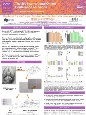
Bisphenol F (BPF) and Bisphenol AF (BPAF) have been used as substitutes for Bisphenol A (BPA), a compound with estrogenic and androgenic properties. This endocrine disruptor compound was already associated with modifying the middle cerebral artery vascular homeostasis by interfering with the nitric oxide pathway. Although BPA has been banned in several countries, recent data suggest that some of its substitutes, such as BPF and BPAF, may pose a risk to human health. However, the available data on their neurovascular effects remain limited. Therefore, the present study evaluated the effects of BPF and BPAF on smooth muscle cells of the middle cerebral artery (SMC-MCA) of Wistar rats. Pure SMC cultures were performed from MCA explants, and MTT assays were performed to assess viability, proliferation, and cytotoxicity. The concentrations tested ranged from 0.0002 to 1000 µM. Planar Cell Surface Area (PCSA) assays were also performed to assess contractile responses to noradrenaline and relaxation responses to sodium nitroprusside at concentrations of 0.002, 0.02, 0.2, 2, and 20 µM. The results showed dose-dependent effects, and both compounds stimulated cell proliferation at low concentrations (0.02 µM) and induced cytotoxicity at high concentrations (200 and 1000 µM), revealing a non-monotonic profile characteristic of endocrine disruptors. Similar to BPA, these compounds modified the vasoactive activity of SMC-MCA, possibly by modulating α1-adrenergic receptors, the NO/sGC pathway, and Ca²⁺ channels, suggesting a potential neurovascular-disrupting effect. BPF altered only the vasorelaxant response at concentrations of 0.002 and 0.02 µM, while BPAF affected both the contractile response (0.002 µM) and the vasorelaxant response (2 µM). It was also noted that, at 0.002 µM, BPAF inhibited SNP-induced relaxation, causing a contractile response. Overall, these findings underscore the need for further studies to fully understand the mechanisms by which BPF and BPAF affect vascular function and the risk of developing neurovascular diseases.
Keywords
Bisphenol F
Bisphenol AF
Bisphenol A
Middle Cerebral Artery
Smooth Muscle Cells
Vasoactive Activity
Neurovascular Toxicology
Poster
Poster_IOCTO2026_Henrique Costa.pdf
Comparative Assessment of Contrast Enhancement Properties of Gadolinium-Based MRI Contrast Agents
Short-term treatment with lithium and its effect on the content of elements and fatty acids in aquatic species of L. minor