Event submissions
Published
This submission belongs to the session S4. Clinical Diagnosis and Prognosis of the event The 1st International Online Conference on Diagnostics
Published date
18 Sep, 2026
Academic Editor
'%3e%3cpath%20d='M12.6657%2010.9104C12.4185%2010.325%2012.0599%209.79327%2011.6097%209.34482C11.1609%208.89508%2010.6293%208.5365%2010.0442%208.28888C10.0389%208.28626%2010.0337%208.28495%2010.0285%208.28233C10.8446%207.69279%2011.3752%206.73249%2011.3752%205.64904C11.3752%203.8542%209.92103%202.39999%208.1262%202.39999C6.33136%202.39999%204.87715%203.8542%204.87715%205.64904C4.87715%206.73249%205.40774%207.69279%206.22393%208.28364C6.21869%208.28626%206.21345%208.28757%206.20821%208.29019C5.62129%208.5378%205.09463%208.89284%204.64265%209.34613C4.1929%209.79493%203.83432%2010.3266%203.58671%2010.9117C3.34345%2011.4845%203.21226%2012.0987%203.20023%2012.7209C3.19988%2012.7349%203.20233%2012.7488%203.20744%2012.7619C3.21255%2012.7749%203.22022%2012.7867%203.22998%2012.7968C3.23975%2012.8068%203.25142%2012.8147%203.26431%2012.8202C3.2772%2012.8256%203.29105%2012.8284%203.30504%2012.8284H4.09109C4.14874%2012.8284%204.19459%2012.7825%204.1959%2012.7262C4.2221%2011.7148%204.62823%2010.7676%205.34617%2010.0497C6.08899%209.30683%207.0755%208.89808%208.1262%208.89808C9.17689%208.89808%2010.1634%209.30683%2010.9062%2010.0497C11.6242%2010.7676%2012.0303%2011.7148%2012.0565%2012.7262C12.0578%2012.7838%2012.1037%2012.8284%2012.1613%2012.8284H12.9474C12.9613%2012.8284%2012.9752%2012.8256%2012.9881%2012.8202C13.001%2012.8147%2013.0126%2012.8068%2013.0224%2012.7968C13.0322%2012.7867%2013.0398%2012.7749%2013.0449%2012.7619C13.0501%2012.7488%2013.0525%2012.7349%2013.0522%2012.7209C13.0391%2012.0947%2012.9094%2011.4855%2012.6657%2010.9104ZM8.1262%207.9024C7.52486%207.9024%206.9589%207.6679%206.53312%207.24211C6.10733%206.81633%205.87283%206.25037%205.87283%205.64904C5.87283%205.0477%206.10733%204.48174%206.53312%204.05596C6.9589%203.63018%207.52486%203.39567%208.1262%203.39567C8.72753%203.39567%209.29349%203.63018%209.71927%204.05596C10.1451%204.48174%2010.3796%205.0477%2010.3796%205.64904C10.3796%206.25037%2010.1451%206.81633%209.71927%207.24211C9.29349%207.6679%208.72753%207.9024%208.1262%207.9024Z'%20fill='%235D1EE1'/%3e%3c/g%3e%3c/svg%3e) Elad Asher
Elad AsherCitation
Yu-Li Wang, Chao-Min Cheng, PACS-Based Radiographic Posture Grid Mapping on a Frontal Spine Radiograph: A Single-Image Proof-of-Concept, in Proceedings of The 1st International Online Conference on Diagnostics, 23 September–24 September 2026, MDPI: Basel, Switzerland
Share
Email
Facebook
Twitter
LinkedIn
PACS-Based Radiographic Posture Grid Mapping on a Frontal Spine Radiograph: A Single-Image Proof-of-Concept
'%3e%3cpath%20d='M12.6647%2010.9104C12.4176%2010.325%2012.0589%209.7933%2011.6088%209.34485C11.16%208.89511%2010.6283%208.53653%2010.0432%208.28891C10.038%208.28629%2010.0327%208.28498%2010.0275%208.28236C10.8437%207.69282%2011.3743%206.73252%2011.3743%205.64907C11.3743%203.85423%209.92005%202.40002%208.12522%202.40002C6.33038%202.40002%204.87618%203.85423%204.87618%205.64907C4.87618%206.73252%205.40677%207.69282%206.22296%208.28367C6.21772%208.28629%206.21248%208.2876%206.20724%208.29022C5.62031%208.53783%205.09365%208.89287%204.64167%209.34616C4.19192%209.79496%203.83334%2010.3266%203.58573%2010.9117C3.34247%2011.4846%203.21128%2012.0987%203.19925%2012.721C3.1989%2012.735%203.20135%2012.7489%203.20646%2012.7619C3.21157%2012.7749%203.21924%2012.7868%203.22901%2012.7968C3.23877%2012.8068%203.25045%2012.8148%203.26334%2012.8202C3.27623%2012.8256%203.29007%2012.8284%203.30406%2012.8284H4.09012C4.14776%2012.8284%204.19362%2012.7825%204.19493%2012.7262C4.22113%2011.7148%204.62726%2010.7676%205.34519%2010.0497C6.08802%209.30686%207.07452%208.89811%208.12522%208.89811C9.17592%208.89811%2010.1624%209.30686%2010.9052%2010.0497C11.6232%2010.7676%2012.0293%2011.7148%2012.0555%2012.7262C12.0568%2012.7839%2012.1027%2012.8284%2012.1603%2012.8284H12.9464C12.9604%2012.8284%2012.9742%2012.8256%2012.9871%2012.8202C13%2012.8148%2013.0117%2012.8068%2013.0214%2012.7968C13.0312%2012.7868%2013.0389%2012.7749%2013.044%2012.7619C13.0491%2012.7489%2013.0515%2012.735%2013.0512%2012.721C13.0381%2012.0947%2012.9084%2011.4856%2012.6647%2010.9104ZM8.12522%207.90243C7.52388%207.90243%206.95792%207.66793%206.53214%207.24215C6.10636%206.81636%205.87185%206.2504%205.87185%205.64907C5.87185%205.04773%206.10636%204.48177%206.53214%204.05599C6.95792%203.63021%207.52388%203.3957%208.12522%203.3957C8.72655%203.3957%209.29252%203.63021%209.7183%204.05599C10.1441%204.48177%2010.3786%205.04773%2010.3786%205.64907C10.3786%206.2504%2010.1441%206.81636%209.7183%207.24215C9.29252%207.66793%208.72655%207.90243%208.12522%207.90243Z'%20fill='%235D1EE1'/%3e%3c/g%3e%3c/svg%3e)
Yu-Li Wang 1,2

Chao-Min Cheng 3
1. Department of Surgery, Hualien Armed Forces General Hospital, Hualien 971, Taiwan
2. International Intercollegiate Ph.D. Program, National Tsing Hua University, Hsinchu 300, Taiwan
3. Institute of Biomedical Engineering, National Tsing Hua University, Hsinchu 300, Taiwan
Abstract
Introduction:
Posture assessment is commonly performed using visual inspection, surface markers, or photographic grids. Although convenient, these methods mainly reflect external body contours and may be influenced by soft-tissue thickness, muscle bulk, clothing, and examiner interpretation. Previous radiographic work has shown that vertebral landmark information can be extracted and standardized for positional comparison. Based on this technical foundation, the present work examined whether skeletal landmarks on a frontal spine radiograph could be displayed on a coordinate grid for posture-oriented visualization.
Methods:
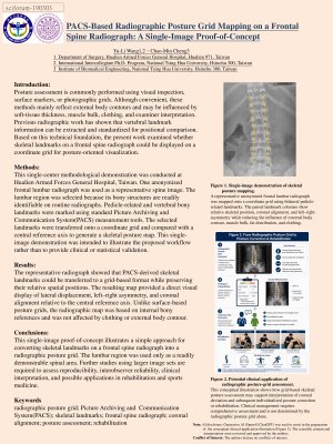
This single-center methodological demonstration was conducted at Hualien Armed Forces General Hospital, Taiwan. One anonymized frontal lumbar radiograph was used as a representative spine image. The lumbar region was selected because its bony structures are readily identifiable on routine radiographs. Pedicle-related and vertebral bony landmarks were marked using standard PACS measurement tools. The selected landmarks were transferred onto a coordinate grid and compared with a central reference axis to generate a skeletal posture map. This single-image demonstration was intended to illustrate the proposed workflow rather than to provide clinical or statistical validation.
Results:
The representative radiograph showed that PACS-derived skeletal landmarks could be transferred to a grid-based format while preserving their relative spatial positions. The resulting map provided a direct visual display of lateral displacement, left–right asymmetry, and coronal alignment relative to the central reference axis. Unlike surface-based posture grids, the radiographic map was based on internal bony references and was not affected by clothing or external body contour.
Conclusions:
This single-image proof-of-concept illustrates a simple approach for converting skeletal landmarks on a frontal spine radiograph into a radiographic posture grid. The lumbar region was used only as a readily demonstrable spinal area. Further studies using larger image sets are required to assess reproducibility, interobserver reliability, clinical interpretation, and possible applications in rehabilitation and sports medicine.
Keywords
Keywords: radiographic posture grid
PACS
skeletal landmarks
frontal spine radiograph
coronal alignment
posture assessment
rehabilitation
Poster
IOCDi2026.pdf
Effect of radiofrequency associated with other interventions on metabolic parameters: a randomized controlled trial
Metformin induces autophagy level changes in heterozygous and CRISPR-edited TSC2 primary fibroblasts