Event submissions
Published
This submission belongs to the session S1. Medical Imaging and Theranostics of the event The 1st International Online Conference on Diagnostics
Published date
18 Sep, 2026
Academic Editor
'%3e%3cpath%20d='M12.6657%2010.9104C12.4185%2010.325%2012.0599%209.79327%2011.6097%209.34482C11.1609%208.89508%2010.6293%208.5365%2010.0442%208.28888C10.0389%208.28626%2010.0337%208.28495%2010.0285%208.28233C10.8446%207.69279%2011.3752%206.73249%2011.3752%205.64904C11.3752%203.8542%209.92103%202.39999%208.1262%202.39999C6.33136%202.39999%204.87715%203.8542%204.87715%205.64904C4.87715%206.73249%205.40774%207.69279%206.22393%208.28364C6.21869%208.28626%206.21345%208.28757%206.20821%208.29019C5.62129%208.5378%205.09463%208.89284%204.64265%209.34613C4.1929%209.79493%203.83432%2010.3266%203.58671%2010.9117C3.34345%2011.4845%203.21226%2012.0987%203.20023%2012.7209C3.19988%2012.7349%203.20233%2012.7488%203.20744%2012.7619C3.21255%2012.7749%203.22022%2012.7867%203.22998%2012.7968C3.23975%2012.8068%203.25142%2012.8147%203.26431%2012.8202C3.2772%2012.8256%203.29105%2012.8284%203.30504%2012.8284H4.09109C4.14874%2012.8284%204.19459%2012.7825%204.1959%2012.7262C4.2221%2011.7148%204.62823%2010.7676%205.34617%2010.0497C6.08899%209.30683%207.0755%208.89808%208.1262%208.89808C9.17689%208.89808%2010.1634%209.30683%2010.9062%2010.0497C11.6242%2010.7676%2012.0303%2011.7148%2012.0565%2012.7262C12.0578%2012.7838%2012.1037%2012.8284%2012.1613%2012.8284H12.9474C12.9613%2012.8284%2012.9752%2012.8256%2012.9881%2012.8202C13.001%2012.8147%2013.0126%2012.8068%2013.0224%2012.7968C13.0322%2012.7867%2013.0398%2012.7749%2013.0449%2012.7619C13.0501%2012.7488%2013.0525%2012.7349%2013.0522%2012.7209C13.0391%2012.0947%2012.9094%2011.4855%2012.6657%2010.9104ZM8.1262%207.9024C7.52486%207.9024%206.9589%207.6679%206.53312%207.24211C6.10733%206.81633%205.87283%206.25037%205.87283%205.64904C5.87283%205.0477%206.10733%204.48174%206.53312%204.05596C6.9589%203.63018%207.52486%203.39567%208.1262%203.39567C8.72753%203.39567%209.29349%203.63018%209.71927%204.05596C10.1451%204.48174%2010.3796%205.0477%2010.3796%205.64904C10.3796%206.25037%2010.1451%206.81633%209.71927%207.24211C9.29349%207.6679%208.72753%207.9024%208.1262%207.9024Z'%20fill='%235D1EE1'/%3e%3c/g%3e%3c/svg%3e) Giorgio Treglia
Giorgio TregliaCitation
Dongxue Liu, Imaging Analysis of Posterior Tibial Tendon Dysfunction: Impact of Accessory Navicular Morphotypes on MRI Indexes, in Proceedings of The 1st International Online Conference on Diagnostics, 23 September–24 September 2026, MDPI: Basel, Switzerland
Share
Email
Facebook
Twitter
LinkedIn
Imaging Analysis of Posterior Tibial Tendon Dysfunction: Impact of Accessory Navicular Morphotypes on MRI Indexes
'%3e%3cpath%20d='M12.6647%2010.9104C12.4176%2010.325%2012.0589%209.7933%2011.6088%209.34485C11.16%208.89511%2010.6283%208.53653%2010.0432%208.28891C10.038%208.28629%2010.0327%208.28498%2010.0275%208.28236C10.8437%207.69282%2011.3743%206.73252%2011.3743%205.64907C11.3743%203.85423%209.92005%202.40002%208.12522%202.40002C6.33038%202.40002%204.87618%203.85423%204.87618%205.64907C4.87618%206.73252%205.40677%207.69282%206.22296%208.28367C6.21772%208.28629%206.21248%208.2876%206.20724%208.29022C5.62031%208.53783%205.09365%208.89287%204.64167%209.34616C4.19192%209.79496%203.83334%2010.3266%203.58573%2010.9117C3.34247%2011.4846%203.21128%2012.0987%203.19925%2012.721C3.1989%2012.735%203.20135%2012.7489%203.20646%2012.7619C3.21157%2012.7749%203.21924%2012.7868%203.22901%2012.7968C3.23877%2012.8068%203.25045%2012.8148%203.26334%2012.8202C3.27623%2012.8256%203.29007%2012.8284%203.30406%2012.8284H4.09012C4.14776%2012.8284%204.19362%2012.7825%204.19493%2012.7262C4.22113%2011.7148%204.62726%2010.7676%205.34519%2010.0497C6.08802%209.30686%207.07452%208.89811%208.12522%208.89811C9.17592%208.89811%2010.1624%209.30686%2010.9052%2010.0497C11.6232%2010.7676%2012.0293%2011.7148%2012.0555%2012.7262C12.0568%2012.7839%2012.1027%2012.8284%2012.1603%2012.8284H12.9464C12.9604%2012.8284%2012.9742%2012.8256%2012.9871%2012.8202C13%2012.8148%2013.0117%2012.8068%2013.0214%2012.7968C13.0312%2012.7868%2013.0389%2012.7749%2013.044%2012.7619C13.0491%2012.7489%2013.0515%2012.735%2013.0512%2012.721C13.0381%2012.0947%2012.9084%2011.4856%2012.6647%2010.9104ZM8.12522%207.90243C7.52388%207.90243%206.95792%207.66793%206.53214%207.24215C6.10636%206.81636%205.87185%206.2504%205.87185%205.64907C5.87185%205.04773%206.10636%204.48177%206.53214%204.05599C6.95792%203.63021%207.52388%203.3957%208.12522%203.3957C8.72655%203.3957%209.29252%203.63021%209.7183%204.05599C10.1441%204.48177%2010.3786%205.04773%2010.3786%205.64907C10.3786%206.2504%2010.1441%206.81636%209.7183%207.24215C9.29252%207.66793%208.72655%207.90243%208.12522%207.90243Z'%20fill='%235D1EE1'/%3e%3c/g%3e%3c/svg%3e)
Dongxue Liu 1
1. Department of Radiology, The Affiliated Hospital of Beihua University, Jilin City, 132000, China
Abstract
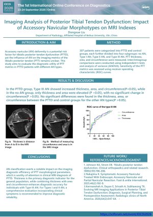
Introduction: Accessory navicular deformity is a potential risk factor for PTTD, yet the influence of accessory navicular (AN) on the imaging metrics of the tibialis posterior tendon (PTT) remains unclear. This study aims to evaluate the diagnostic utility of PTT metrics in tibialis posterior tendon dysfunction (PTTD) patients with different AN types.
Methods:307 patients were categorized into PTTD and control groups, each further divided into four subgroups: no AN, Type I AN, Type II AN, and Type III AN. PTT thickness, area, and circumference were measured. Inter/intragroup comparisons were conducted using independent t-tests and analysis of variance (ANOVA). Sensitivity of the PTT metrics was assessed using receiver operating characteristic (ROC) curves.
Results:In the PTTD group, Type III AN showed increased thickness, area, and circumference (P < 0.05), while in the no-accessory navicular group, only thickness and area were elevated (P < 0.05), with no significant change in circumference (P > 0.05). No significant differences were found in the thickness, area, or circumference between the posterior tibial tendon dysfunction and control groups for the other accessory navicular types (P > 0.05).
Conclusion: Accessory navicular classification exerts a notable impact on the imaging diagnostic efficiency of PTT morphological parameters, which is worthy of attention in clinical MRI diagnosis of PTTD. Thickness is the primary diagnostic indicator for the general population, while combining thickness with area and circumference enhances diagnostic accuracy for individuals with type III AN. For types I and II AN, a comprehensive evaluation incorporating clinical symptoms is recommended to improve diagnostic reliability
Keywords
Accessory navicular
Posterior tibial tendon
Posterior tibial tendon dysfunction.
Poster
会议摘要模版.pdf
Prevalence of Hypertension and Diabetes Among End-Stage Renal Disease Patients on Dialysis at the Renal Dialysis Center in Hodeidah, Yemen
From Diagnostic Imaging to Systemic Prevention: An Integrated CEM–AI Framework for Oncologic, Osteoporotic, and Vascular Risk Stratification