Event submissions
Published
This submission belongs to the session S3. Pediatric Dentistry and Orthodontics of the event The 1st International Online Conference on Dentistry
Published date
02 Oct, 2026
Academic Editor
'%3e%3cpath%20d='M12.6657%2010.9104C12.4185%2010.325%2012.0599%209.79327%2011.6097%209.34482C11.1609%208.89508%2010.6293%208.5365%2010.0442%208.28888C10.0389%208.28626%2010.0337%208.28495%2010.0285%208.28233C10.8446%207.69279%2011.3752%206.73249%2011.3752%205.64904C11.3752%203.8542%209.92103%202.39999%208.1262%202.39999C6.33136%202.39999%204.87715%203.8542%204.87715%205.64904C4.87715%206.73249%205.40774%207.69279%206.22393%208.28364C6.21869%208.28626%206.21345%208.28757%206.20821%208.29019C5.62129%208.5378%205.09463%208.89284%204.64265%209.34613C4.1929%209.79493%203.83432%2010.3266%203.58671%2010.9117C3.34345%2011.4845%203.21226%2012.0987%203.20023%2012.7209C3.19988%2012.7349%203.20233%2012.7488%203.20744%2012.7619C3.21255%2012.7749%203.22022%2012.7867%203.22998%2012.7968C3.23975%2012.8068%203.25142%2012.8147%203.26431%2012.8202C3.2772%2012.8256%203.29105%2012.8284%203.30504%2012.8284H4.09109C4.14874%2012.8284%204.19459%2012.7825%204.1959%2012.7262C4.2221%2011.7148%204.62823%2010.7676%205.34617%2010.0497C6.08899%209.30683%207.0755%208.89808%208.1262%208.89808C9.17689%208.89808%2010.1634%209.30683%2010.9062%2010.0497C11.6242%2010.7676%2012.0303%2011.7148%2012.0565%2012.7262C12.0578%2012.7838%2012.1037%2012.8284%2012.1613%2012.8284H12.9474C12.9613%2012.8284%2012.9752%2012.8256%2012.9881%2012.8202C13.001%2012.8147%2013.0126%2012.8068%2013.0224%2012.7968C13.0322%2012.7867%2013.0398%2012.7749%2013.0449%2012.7619C13.0501%2012.7488%2013.0525%2012.7349%2013.0522%2012.7209C13.0391%2012.0947%2012.9094%2011.4855%2012.6657%2010.9104ZM8.1262%207.9024C7.52486%207.9024%206.9589%207.6679%206.53312%207.24211C6.10733%206.81633%205.87283%206.25037%205.87283%205.64904C5.87283%205.0477%206.10733%204.48174%206.53312%204.05596C6.9589%203.63018%207.52486%203.39567%208.1262%203.39567C8.72753%203.39567%209.29349%203.63018%209.71927%204.05596C10.1451%204.48174%2010.3796%205.0477%2010.3796%205.64904C10.3796%206.25037%2010.1451%206.81633%209.71927%207.24211C9.29349%207.6679%208.72753%207.9024%208.1262%207.9024Z'%20fill='%235D1EE1'/%3e%3c/g%3e%3c/svg%3e) Georgios Romanos
Georgios RomanosCitation
Ralitsa Bogovska-Gigova, Krasimir Hristov, Victoria Zlateva, Micro-CT analysis of enamel mineral density in erupted, impacted and unerupted immature third molars, in Proceedings of The 1st International Online Conference on Dentistry, 7 October–9 October 2026, MDPI: Basel, Switzerland
Share
Email
Facebook
Twitter
LinkedIn
Micro-CT analysis of enamel mineral density in erupted, impacted and unerupted immature third molars
'%3e%3cpath%20d='M12.6647%2010.9104C12.4176%2010.325%2012.0589%209.7933%2011.6088%209.34485C11.16%208.89511%2010.6283%208.53653%2010.0432%208.28891C10.038%208.28629%2010.0327%208.28498%2010.0275%208.28236C10.8437%207.69282%2011.3743%206.73252%2011.3743%205.64907C11.3743%203.85423%209.92005%202.40002%208.12522%202.40002C6.33038%202.40002%204.87618%203.85423%204.87618%205.64907C4.87618%206.73252%205.40677%207.69282%206.22296%208.28367C6.21772%208.28629%206.21248%208.2876%206.20724%208.29022C5.62031%208.53783%205.09365%208.89287%204.64167%209.34616C4.19192%209.79496%203.83334%2010.3266%203.58573%2010.9117C3.34247%2011.4846%203.21128%2012.0987%203.19925%2012.721C3.1989%2012.735%203.20135%2012.7489%203.20646%2012.7619C3.21157%2012.7749%203.21924%2012.7868%203.22901%2012.7968C3.23877%2012.8068%203.25045%2012.8148%203.26334%2012.8202C3.27623%2012.8256%203.29007%2012.8284%203.30406%2012.8284H4.09012C4.14776%2012.8284%204.19362%2012.7825%204.19493%2012.7262C4.22113%2011.7148%204.62726%2010.7676%205.34519%2010.0497C6.08802%209.30686%207.07452%208.89811%208.12522%208.89811C9.17592%208.89811%2010.1624%209.30686%2010.9052%2010.0497C11.6232%2010.7676%2012.0293%2011.7148%2012.0555%2012.7262C12.0568%2012.7839%2012.1027%2012.8284%2012.1603%2012.8284H12.9464C12.9604%2012.8284%2012.9742%2012.8256%2012.9871%2012.8202C13%2012.8148%2013.0117%2012.8068%2013.0214%2012.7968C13.0312%2012.7868%2013.0389%2012.7749%2013.044%2012.7619C13.0491%2012.7489%2013.0515%2012.735%2013.0512%2012.721C13.0381%2012.0947%2012.9084%2011.4856%2012.6647%2010.9104ZM8.12522%207.90243C7.52388%207.90243%206.95792%207.66793%206.53214%207.24215C6.10636%206.81636%205.87185%206.2504%205.87185%205.64907C5.87185%205.04773%206.10636%204.48177%206.53214%204.05599C6.95792%203.63021%207.52388%203.3957%208.12522%203.3957C8.72655%203.3957%209.29252%203.63021%209.7183%204.05599C10.1441%204.48177%2010.3786%205.04773%2010.3786%205.64907C10.3786%206.2504%2010.1441%206.81636%209.7183%207.24215C9.29252%207.66793%208.72655%207.90243%208.12522%207.90243Z'%20fill='%235D1EE1'/%3e%3c/g%3e%3c/svg%3e)
Ralitsa Bogovska-Gigova 1
Krasimir Hristov 1
Victoria Zlateva 1
1. Medical University Sofia, Faculty of Dental Medicine, department of pediatrics dentistry, Sofia, Bulgaria
Abstract
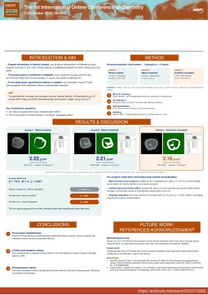
Background: Enamel mineral density reflects the degree of tissue maturation and strongly influences the mechanical properties of teeth. Because third molars complete their development over a broad age range, they represent a suitable model for investigating enamel mineralization at different developmental stages. This study compared enamel mineral density among erupted, impaccted, and unerupted immature third molars using micro-computed tomography (micro-CT) calibrated with a hydroxyapatite phantom.
Methods: Thirty-nine extracted human third molars were allocated into three groups (n = 13 per group): mature erupted, mature unerupted (impacted), and immature unerupted. All specimens were scanned using micro-computed tomography (micro-CT). Hydroxyapatite calibration phantoms with mineral densities of 0.30 and 1.25 g/cm³ were scanned using identical parameters to establish a linear calibration curve between grayscale values and mineral density. Enamel mineral density (MD, g/cm³) was subsequently calculated using CTAn software. Median enamel mineral density was compared among groups with a statistival significance set at p<0.05.
Results: Enamel mineral density differed significantly among the three groups (p<0.001). Median mineral density was 4.03 g/cm³ for mature erupted teeth, 4.02 g/cm³ for mature unerupted teeth, and 3.92 g/cm³ for immature unerupted teeth. Post hoc analysis demonstrated no significant difference between mature erupted and mature unerupted teeth. In contrast, immature unerupted teeth exhibited significantly lower enamel mineral density than both mature erupted teeth and mature unerupted teeth.
Conclusions: Mature erupted and mature unerupted third molars showed comparable density, while immature unerupted teeth had significantly lower mineralization, indicating that key stages of enamel mineralization are completed pre-eruptively. Post-eruptive maturation appears to have minimal effect on bulk enamel mineral density as measured by micro-CT.
Keywords
enamel mineral density
enamel maturation
micro-computed tomography
dental enamel.
Poster
Poster IOCDT2026_Poster_Micro-CT_third_molars_1.pdf
One-Stage Digital Rehabilitation of a Severely Atrophic Posterior Maxilla Using a Customized Zirconia Mesh, Lateral Sinus Floor Elevation, and Simultaneous Implant Placement: A Clinical Case Report
Surface defect burden as determinant of cyclic fatigue fragmentation in counterfeit endodontic files: SEM-based comparative study