Event submissions
Published
This submission belongs to the session S7. Digital Dentistry of the event The 1st International Online Conference on Dentistry
Published date
02 Oct, 2026
Academic Editor
'%3e%3cpath%20d='M12.6657%2010.9104C12.4185%2010.325%2012.0599%209.79327%2011.6097%209.34482C11.1609%208.89508%2010.6293%208.5365%2010.0442%208.28888C10.0389%208.28626%2010.0337%208.28495%2010.0285%208.28233C10.8446%207.69279%2011.3752%206.73249%2011.3752%205.64904C11.3752%203.8542%209.92103%202.39999%208.1262%202.39999C6.33136%202.39999%204.87715%203.8542%204.87715%205.64904C4.87715%206.73249%205.40774%207.69279%206.22393%208.28364C6.21869%208.28626%206.21345%208.28757%206.20821%208.29019C5.62129%208.5378%205.09463%208.89284%204.64265%209.34613C4.1929%209.79493%203.83432%2010.3266%203.58671%2010.9117C3.34345%2011.4845%203.21226%2012.0987%203.20023%2012.7209C3.19988%2012.7349%203.20233%2012.7488%203.20744%2012.7619C3.21255%2012.7749%203.22022%2012.7867%203.22998%2012.7968C3.23975%2012.8068%203.25142%2012.8147%203.26431%2012.8202C3.2772%2012.8256%203.29105%2012.8284%203.30504%2012.8284H4.09109C4.14874%2012.8284%204.19459%2012.7825%204.1959%2012.7262C4.2221%2011.7148%204.62823%2010.7676%205.34617%2010.0497C6.08899%209.30683%207.0755%208.89808%208.1262%208.89808C9.17689%208.89808%2010.1634%209.30683%2010.9062%2010.0497C11.6242%2010.7676%2012.0303%2011.7148%2012.0565%2012.7262C12.0578%2012.7838%2012.1037%2012.8284%2012.1613%2012.8284H12.9474C12.9613%2012.8284%2012.9752%2012.8256%2012.9881%2012.8202C13.001%2012.8147%2013.0126%2012.8068%2013.0224%2012.7968C13.0322%2012.7867%2013.0398%2012.7749%2013.0449%2012.7619C13.0501%2012.7488%2013.0525%2012.7349%2013.0522%2012.7209C13.0391%2012.0947%2012.9094%2011.4855%2012.6657%2010.9104ZM8.1262%207.9024C7.52486%207.9024%206.9589%207.6679%206.53312%207.24211C6.10733%206.81633%205.87283%206.25037%205.87283%205.64904C5.87283%205.0477%206.10733%204.48174%206.53312%204.05596C6.9589%203.63018%207.52486%203.39567%208.1262%203.39567C8.72753%203.39567%209.29349%203.63018%209.71927%204.05596C10.1451%204.48174%2010.3796%205.0477%2010.3796%205.64904C10.3796%206.25037%2010.1451%206.81633%209.71927%207.24211C9.29349%207.6679%208.72753%207.9024%208.1262%207.9024Z'%20fill='%235D1EE1'/%3e%3c/g%3e%3c/svg%3e) Claude Jaquiéry
Claude JaquiéryCitation
Tirhab Eltayeb, Amr Solaiman, Khaled Borghol, Virtual Surgical Planning for Removal of an Ectopic Maxillary Third Molar Associated with a Large Maxillary Sinus Cyst: A Case Report, in Proceedings of The 1st International Online Conference on Dentistry, 7 October–9 October 2026, MDPI: Basel, Switzerland
Share
Email
Facebook
Twitter
LinkedIn
Virtual Surgical Planning for Removal of an Ectopic Maxillary Third Molar Associated with a Large Maxillary Sinus Cyst: A Case Report
'%3e%3cpath%20d='M12.6647%2010.9104C12.4176%2010.325%2012.0589%209.7933%2011.6088%209.34485C11.16%208.89511%2010.6283%208.53653%2010.0432%208.28891C10.038%208.28629%2010.0327%208.28498%2010.0275%208.28236C10.8437%207.69282%2011.3743%206.73252%2011.3743%205.64907C11.3743%203.85423%209.92005%202.40002%208.12522%202.40002C6.33038%202.40002%204.87618%203.85423%204.87618%205.64907C4.87618%206.73252%205.40677%207.69282%206.22296%208.28367C6.21772%208.28629%206.21248%208.2876%206.20724%208.29022C5.62031%208.53783%205.09365%208.89287%204.64167%209.34616C4.19192%209.79496%203.83334%2010.3266%203.58573%2010.9117C3.34247%2011.4846%203.21128%2012.0987%203.19925%2012.721C3.1989%2012.735%203.20135%2012.7489%203.20646%2012.7619C3.21157%2012.7749%203.21924%2012.7868%203.22901%2012.7968C3.23877%2012.8068%203.25045%2012.8148%203.26334%2012.8202C3.27623%2012.8256%203.29007%2012.8284%203.30406%2012.8284H4.09012C4.14776%2012.8284%204.19362%2012.7825%204.19493%2012.7262C4.22113%2011.7148%204.62726%2010.7676%205.34519%2010.0497C6.08802%209.30686%207.07452%208.89811%208.12522%208.89811C9.17592%208.89811%2010.1624%209.30686%2010.9052%2010.0497C11.6232%2010.7676%2012.0293%2011.7148%2012.0555%2012.7262C12.0568%2012.7839%2012.1027%2012.8284%2012.1603%2012.8284H12.9464C12.9604%2012.8284%2012.9742%2012.8256%2012.9871%2012.8202C13%2012.8148%2013.0117%2012.8068%2013.0214%2012.7968C13.0312%2012.7868%2013.0389%2012.7749%2013.044%2012.7619C13.0491%2012.7489%2013.0515%2012.735%2013.0512%2012.721C13.0381%2012.0947%2012.9084%2011.4856%2012.6647%2010.9104ZM8.12522%207.90243C7.52388%207.90243%206.95792%207.66793%206.53214%207.24215C6.10636%206.81636%205.87185%206.2504%205.87185%205.64907C5.87185%205.04773%206.10636%204.48177%206.53214%204.05599C6.95792%203.63021%207.52388%203.3957%208.12522%203.3957C8.72655%203.3957%209.29252%203.63021%209.7183%204.05599C10.1441%204.48177%2010.3786%205.04773%2010.3786%205.64907C10.3786%206.2504%2010.1441%206.81636%209.7183%207.24215C9.29252%207.66793%208.72655%207.90243%208.12522%207.90243Z'%20fill='%235D1EE1'/%3e%3c/g%3e%3c/svg%3e)
Tirhab Eltayeb 1
Amr Solaiman 1
Khaled Borghol 1
1. Department of Oral and Maxillofacial Surgery, The Shrewsbury and Telford Hospital NHS Trust, Shrewsbury, UK
Abstract
Abstract
Introduction
Ectopic eruption of maxillary third molars into the maxillary sinus is rare and often associated with cystic lesions and non-specific sinonasal symptoms. Delayed diagnosis may result in progressive sinus expansion and complications.
Case Presentation
A 37-year-old female smoker presented with purulent discharge distal to the upper right second molar (UR7), persistent bad taste, right-sided nasal obstruction, and recurrent sinusitis symptoms.
A CT scan in 2020 for headaches showed a partially visualised expansile right maxillary sinus lesion, with no further follow-up. A recent radiograph demonstrated a large radiopaque lesion occupying most of the right maxillary sinus, associated with an ectopic maxillary third molar and distal bone loss adjacent to UR7. CT confirmed a well-defined cystic lesion associated with the ectopic tooth.
Management
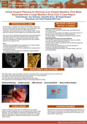
Pre-operative intraoral scanning of the maxillary and mandibular arches was performed to facilitate virtual surgical planning (VSP) and fabrication of a patient-specific surgical guide. The ectopic upper right third molar was removed via an intraoral approach using guided access. The associated cystic lesion was enucleated concurrently, and the resulting oroantral communication was closed primarily. Following removal of the ectopic tooth and associated pathology, the bony window was repositioned and stabilised using fixation plates.
Post-operative management included clinical and radiographic follow-up, with histopathological examination of the excised lesion.
Discussion
Ectopic third molars within the maxillary sinus are uncommon and frequently associated with dentigerous cysts. This case highlights the importance of follow-up of incidental radiographic findings and the role of advanced imaging in diagnosis and surgical planning. Early intervention may prevent extensive sinus involvement.
Conclusion
This case highlights the value of virtual surgical planning and a patient-specific surgical guide in improving localisation and surgical precision for ectopic maxillary third molar removal.
Keywords
Ectopic third molar
maxillary sinus
virtual surgical planning
digital dentistry
guided surgery
dentigerous cyst
patient-specific surgical guide
intraoral scanning
oral surgery
maxillofacial surgery
Poster
IOCDT2026-Poster-TE.pdf
Composite Dental Material Design and Testing Using Mechanical Engineering Methods
Patient Attitudes Toward AI in Periodontal Health Promotion and Assessment