Event submissions
Published
This submission belongs to the session S6. Lasers in Dentistry of the event The 1st International Online Conference on Dentistry
Published date
02 Oct, 2026
Academic Editor
'%3e%3cpath%20d='M12.6657%2010.9104C12.4185%2010.325%2012.0599%209.79327%2011.6097%209.34482C11.1609%208.89508%2010.6293%208.5365%2010.0442%208.28888C10.0389%208.28626%2010.0337%208.28495%2010.0285%208.28233C10.8446%207.69279%2011.3752%206.73249%2011.3752%205.64904C11.3752%203.8542%209.92103%202.39999%208.1262%202.39999C6.33136%202.39999%204.87715%203.8542%204.87715%205.64904C4.87715%206.73249%205.40774%207.69279%206.22393%208.28364C6.21869%208.28626%206.21345%208.28757%206.20821%208.29019C5.62129%208.5378%205.09463%208.89284%204.64265%209.34613C4.1929%209.79493%203.83432%2010.3266%203.58671%2010.9117C3.34345%2011.4845%203.21226%2012.0987%203.20023%2012.7209C3.19988%2012.7349%203.20233%2012.7488%203.20744%2012.7619C3.21255%2012.7749%203.22022%2012.7867%203.22998%2012.7968C3.23975%2012.8068%203.25142%2012.8147%203.26431%2012.8202C3.2772%2012.8256%203.29105%2012.8284%203.30504%2012.8284H4.09109C4.14874%2012.8284%204.19459%2012.7825%204.1959%2012.7262C4.2221%2011.7148%204.62823%2010.7676%205.34617%2010.0497C6.08899%209.30683%207.0755%208.89808%208.1262%208.89808C9.17689%208.89808%2010.1634%209.30683%2010.9062%2010.0497C11.6242%2010.7676%2012.0303%2011.7148%2012.0565%2012.7262C12.0578%2012.7838%2012.1037%2012.8284%2012.1613%2012.8284H12.9474C12.9613%2012.8284%2012.9752%2012.8256%2012.9881%2012.8202C13.001%2012.8147%2013.0126%2012.8068%2013.0224%2012.7968C13.0322%2012.7867%2013.0398%2012.7749%2013.0449%2012.7619C13.0501%2012.7488%2013.0525%2012.7349%2013.0522%2012.7209C13.0391%2012.0947%2012.9094%2011.4855%2012.6657%2010.9104ZM8.1262%207.9024C7.52486%207.9024%206.9589%207.6679%206.53312%207.24211C6.10733%206.81633%205.87283%206.25037%205.87283%205.64904C5.87283%205.0477%206.10733%204.48174%206.53312%204.05596C6.9589%203.63018%207.52486%203.39567%208.1262%203.39567C8.72753%203.39567%209.29349%203.63018%209.71927%204.05596C10.1451%204.48174%2010.3796%205.0477%2010.3796%205.64904C10.3796%206.25037%2010.1451%206.81633%209.71927%207.24211C9.29349%207.6679%208.72753%207.9024%208.1262%207.9024Z'%20fill='%235D1EE1'/%3e%3c/g%3e%3c/svg%3e) Samir Nammour
Samir NammourCitation
Kakhramon Shomurodov, Rano Mirkhusanova, Effect of Laser Patterned Microcoagulation on Outcomes of Connective Tissue Graft at Implant Sites, in Proceedings of The 1st International Online Conference on Dentistry, 7 October–9 October 2026, MDPI: Basel, Switzerland
Share
Email
Facebook
Twitter
LinkedIn
Effect of Laser Patterned Microcoagulation on Outcomes of Connective Tissue Graft at Implant Sites
'%3e%3cpath%20d='M12.6647%2010.9104C12.4176%2010.325%2012.0589%209.7933%2011.6088%209.34485C11.16%208.89511%2010.6283%208.53653%2010.0432%208.28891C10.038%208.28629%2010.0327%208.28498%2010.0275%208.28236C10.8437%207.69282%2011.3743%206.73252%2011.3743%205.64907C11.3743%203.85423%209.92005%202.40002%208.12522%202.40002C6.33038%202.40002%204.87618%203.85423%204.87618%205.64907C4.87618%206.73252%205.40677%207.69282%206.22296%208.28367C6.21772%208.28629%206.21248%208.2876%206.20724%208.29022C5.62031%208.53783%205.09365%208.89287%204.64167%209.34616C4.19192%209.79496%203.83334%2010.3266%203.58573%2010.9117C3.34247%2011.4846%203.21128%2012.0987%203.19925%2012.721C3.1989%2012.735%203.20135%2012.7489%203.20646%2012.7619C3.21157%2012.7749%203.21924%2012.7868%203.22901%2012.7968C3.23877%2012.8068%203.25045%2012.8148%203.26334%2012.8202C3.27623%2012.8256%203.29007%2012.8284%203.30406%2012.8284H4.09012C4.14776%2012.8284%204.19362%2012.7825%204.19493%2012.7262C4.22113%2011.7148%204.62726%2010.7676%205.34519%2010.0497C6.08802%209.30686%207.07452%208.89811%208.12522%208.89811C9.17592%208.89811%2010.1624%209.30686%2010.9052%2010.0497C11.6232%2010.7676%2012.0293%2011.7148%2012.0555%2012.7262C12.0568%2012.7839%2012.1027%2012.8284%2012.1603%2012.8284H12.9464C12.9604%2012.8284%2012.9742%2012.8256%2012.9871%2012.8202C13%2012.8148%2013.0117%2012.8068%2013.0214%2012.7968C13.0312%2012.7868%2013.0389%2012.7749%2013.044%2012.7619C13.0491%2012.7489%2013.0515%2012.735%2013.0512%2012.721C13.0381%2012.0947%2012.9084%2011.4856%2012.6647%2010.9104ZM8.12522%207.90243C7.52388%207.90243%206.95792%207.66793%206.53214%207.24215C6.10636%206.81636%205.87185%206.2504%205.87185%205.64907C5.87185%205.04773%206.10636%204.48177%206.53214%204.05599C6.95792%203.63021%207.52388%203.3957%208.12522%203.3957C8.72655%203.3957%209.29252%203.63021%209.7183%204.05599C10.1441%204.48177%2010.3786%205.04773%2010.3786%205.64907C10.3786%206.2504%2010.1441%206.81636%209.7183%207.24215C9.29252%207.66793%208.72655%207.90243%208.12522%207.90243Z'%20fill='%235D1EE1'/%3e%3c/g%3e%3c/svg%3e)
Kakhramon Shomurodov 1
Rano Mirkhusanova 1
1. Department of Maxillofacial Surgery, Tashkent State Medical University, Tashkent, 100109, Uzbekistan
Abstract
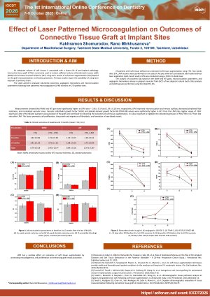
Introduction. An adequate volume of soft tissues is associated with a lower risk of peri-implant pathology. Connective tissue graft (CTG) is commonly used to restore sufficient volume of keratinized mucosa width (KMW) and increase mucosal thickness (MT). Long-term results of soft tissue augmentation (STA) depend on the graft revascularization and healing processes. Previous studies report the possibility of using laser exposure to enhance them. The study aimed to evaluate volumetric outcomes, angiogenic biomarkers and microcirculation parameters following laser patterned microcoagulation (LPM) sessions on CTG-grafted sites. Methods. 25 patients with soft tissue dehiscence underwent soft tissue augmentation using CTG. Two weeks after STA, LPM sessions were performed on one side of the jaw, while the contralateral side healed without laser application (split-mouth study). LPM was conducted using a 1550 nm diode laser. The analysis of outcomes was based on the KMW and MT gains, microcirculation parameters, and angiogenic biomarkers release in gingival crevicular fluid (GCF) of two adjacent natural teeth. Data analysis and plotting were performed using the OriginPro 8.6. Results. Measurements showed that KMW and MT gain were significantly higher at LPM sites – 1.96 ± 0.14 and 1.34 ± 0.18 mm, respectively. LPM improved microcirculation and venous outflow, decreased peripheral flow resistance, and normalized vascular tonus. Vascular endothelial growth factor (VEGF) and platelet-derived growth factor-BB (PDGF-BB) values were significantly higher in GCF from the LPM sites. Conclusions. LPM has a positive effect on outcomes of soft tissue augmentation by promoting neocollagenesis, cell proliferation and enhancing graft revascularization.
Keywords
Diode laser
Soft tissue augmentation
Microcirculation
Biomarker
Dental implantation
Poster
IOCDT2026-Poster Shomurodov, Mirkhusanova.pdf
COMPARATIVE ASSESSMENT OF BOLTON RATIO MEASUREMENTS ON PLASTER AND DIGITAL DENTAL MODELS USING DIFFERENT MEASUREMENT TECHNIQUES
Effect of electronic cigarettes on optical properties of long-lasting composites for CAD/CAM systems