Event submissions
Published
This submission belongs to the session S4. Restorative Dentistry, Endodontics, and Dental Traumatology of the event The 1st International Online Conference on Dentistry
Published date
02 Oct, 2026
Academic Editor
'%3e%3cpath%20d='M12.6657%2010.9104C12.4185%2010.325%2012.0599%209.79327%2011.6097%209.34482C11.1609%208.89508%2010.6293%208.5365%2010.0442%208.28888C10.0389%208.28626%2010.0337%208.28495%2010.0285%208.28233C10.8446%207.69279%2011.3752%206.73249%2011.3752%205.64904C11.3752%203.8542%209.92103%202.39999%208.1262%202.39999C6.33136%202.39999%204.87715%203.8542%204.87715%205.64904C4.87715%206.73249%205.40774%207.69279%206.22393%208.28364C6.21869%208.28626%206.21345%208.28757%206.20821%208.29019C5.62129%208.5378%205.09463%208.89284%204.64265%209.34613C4.1929%209.79493%203.83432%2010.3266%203.58671%2010.9117C3.34345%2011.4845%203.21226%2012.0987%203.20023%2012.7209C3.19988%2012.7349%203.20233%2012.7488%203.20744%2012.7619C3.21255%2012.7749%203.22022%2012.7867%203.22998%2012.7968C3.23975%2012.8068%203.25142%2012.8147%203.26431%2012.8202C3.2772%2012.8256%203.29105%2012.8284%203.30504%2012.8284H4.09109C4.14874%2012.8284%204.19459%2012.7825%204.1959%2012.7262C4.2221%2011.7148%204.62823%2010.7676%205.34617%2010.0497C6.08899%209.30683%207.0755%208.89808%208.1262%208.89808C9.17689%208.89808%2010.1634%209.30683%2010.9062%2010.0497C11.6242%2010.7676%2012.0303%2011.7148%2012.0565%2012.7262C12.0578%2012.7838%2012.1037%2012.8284%2012.1613%2012.8284H12.9474C12.9613%2012.8284%2012.9752%2012.8256%2012.9881%2012.8202C13.001%2012.8147%2013.0126%2012.8068%2013.0224%2012.7968C13.0322%2012.7867%2013.0398%2012.7749%2013.0449%2012.7619C13.0501%2012.7488%2013.0525%2012.7349%2013.0522%2012.7209C13.0391%2012.0947%2012.9094%2011.4855%2012.6657%2010.9104ZM8.1262%207.9024C7.52486%207.9024%206.9589%207.6679%206.53312%207.24211C6.10733%206.81633%205.87283%206.25037%205.87283%205.64904C5.87283%205.0477%206.10733%204.48174%206.53312%204.05596C6.9589%203.63018%207.52486%203.39567%208.1262%203.39567C8.72753%203.39567%209.29349%203.63018%209.71927%204.05596C10.1451%204.48174%2010.3796%205.0477%2010.3796%205.64904C10.3796%206.25037%2010.1451%206.81633%209.71927%207.24211C9.29349%207.6679%208.72753%207.9024%208.1262%207.9024Z'%20fill='%235D1EE1'/%3e%3c/g%3e%3c/svg%3e) Gianrico Spagnuolo
Gianrico SpagnuoloCitation
Chahhat ., Artificial Intelligence in Endodontic Radiology: Current Status of AI Models for CBCT-Based Periapical Pathosis Diagnosis, in Proceedings of The 1st International Online Conference on Dentistry, 7 October–9 October 2026, MDPI: Basel, Switzerland
Share
Email
Facebook
Twitter
LinkedIn
Artificial Intelligence in Endodontic Radiology: Current Status of AI Models for CBCT-Based Periapical Pathosis Diagnosis
'%3e%3cpath%20d='M12.6647%2010.9104C12.4176%2010.325%2012.0589%209.7933%2011.6088%209.34485C11.16%208.89511%2010.6283%208.53653%2010.0432%208.28891C10.038%208.28629%2010.0327%208.28498%2010.0275%208.28236C10.8437%207.69282%2011.3743%206.73252%2011.3743%205.64907C11.3743%203.85423%209.92005%202.40002%208.12522%202.40002C6.33038%202.40002%204.87618%203.85423%204.87618%205.64907C4.87618%206.73252%205.40677%207.69282%206.22296%208.28367C6.21772%208.28629%206.21248%208.2876%206.20724%208.29022C5.62031%208.53783%205.09365%208.89287%204.64167%209.34616C4.19192%209.79496%203.83334%2010.3266%203.58573%2010.9117C3.34247%2011.4846%203.21128%2012.0987%203.19925%2012.721C3.1989%2012.735%203.20135%2012.7489%203.20646%2012.7619C3.21157%2012.7749%203.21924%2012.7868%203.22901%2012.7968C3.23877%2012.8068%203.25045%2012.8148%203.26334%2012.8202C3.27623%2012.8256%203.29007%2012.8284%203.30406%2012.8284H4.09012C4.14776%2012.8284%204.19362%2012.7825%204.19493%2012.7262C4.22113%2011.7148%204.62726%2010.7676%205.34519%2010.0497C6.08802%209.30686%207.07452%208.89811%208.12522%208.89811C9.17592%208.89811%2010.1624%209.30686%2010.9052%2010.0497C11.6232%2010.7676%2012.0293%2011.7148%2012.0555%2012.7262C12.0568%2012.7839%2012.1027%2012.8284%2012.1603%2012.8284H12.9464C12.9604%2012.8284%2012.9742%2012.8256%2012.9871%2012.8202C13%2012.8148%2013.0117%2012.8068%2013.0214%2012.7968C13.0312%2012.7868%2013.0389%2012.7749%2013.044%2012.7619C13.0491%2012.7489%2013.0515%2012.735%2013.0512%2012.721C13.0381%2012.0947%2012.9084%2011.4856%2012.6647%2010.9104ZM8.12522%207.90243C7.52388%207.90243%206.95792%207.66793%206.53214%207.24215C6.10636%206.81636%205.87185%206.2504%205.87185%205.64907C5.87185%205.04773%206.10636%204.48177%206.53214%204.05599C6.95792%203.63021%207.52388%203.3957%208.12522%203.3957C8.72655%203.3957%209.29252%203.63021%209.7183%204.05599C10.1441%204.48177%2010.3786%205.04773%2010.3786%205.64907C10.3786%206.2504%2010.1441%206.81636%209.7183%207.24215C9.29252%207.66793%208.72655%207.90243%208.12522%207.90243Z'%20fill='%235D1EE1'/%3e%3c/g%3e%3c/svg%3e)
Chahhat . 1
1. Department of Conservative Dentistry and Endodontics, Punjab Government Dental College and Hospital, Baba Farid University of Health Sciences, Amritsar, 143001
Abstract
Background:
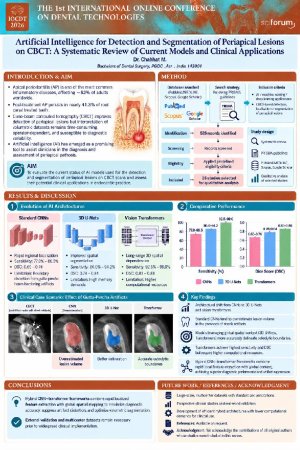
Apical periodontitis (AP) is one of the most common inflammatory diseases affecting the tissues surrounding the root apex and remains a significant global oral health problem. Studies have reported that AP affects approximately 52% of adults worldwide, while post-treatment apical periodontitis persists in nearly 41.3% of root canal treated teeth. Cone-beam computed tomography (CBCT) has improved the detection of periapical lesions compared with conventional radiography, interpretation of volumetric datasets remains time-consuming, operator-dependent, and susceptible to diagnostic variability. Artificial intelligence (AI) has emerged as a promising tool to assist clinicians in the diagnosis and assessment of periapical pathosis.
Aim:
To evaluate the current status of AI models used for the detection and segmentation of periapical lesions on CBCT scans and assess their potential clinical applications in endodontic practice.
Methods:
A systematic literature search was conducted following PRISMA guidelines using PubMed/MEDLINE, Scopus, and Google Scholar. Studies investigating AI, machine learning, or deep learning applications for CBCT-based detection, localization, or segmentation of periapical lesions were included. After screening 526 records and applying predefined eligibility criteria, 26 studies were selected for qualitative analysis.
Results:
The reviewed studies demonstrated an architectural shift from CNNs to 3D U-Nets and vision transformers. Standard CNNs offer rapid regional localization (sensitivity: 77.8%–88.5%, DSC: 0.65–0.74) but suffer boundary distortion from gutta-percha beam-hardening artifacts. 3D U-Nets improve spatial segmentation (sensitivity: 86.0%–94.2%, DSC: 0.78–0.84) despite high memory demands. Transformers model long-range 3D spatial dependencies (sensitivity: 92.5%–99.0%, DSC: 0.83–0.88), though require higher computational resources. In comparative clinical evaluations of root-filled molars with streak artifacts, standard CNNs overestimated lesion volume, whereas models leveraging global spatial context accurately delineated osteolytic boundaries.
Conclusion:
Hybrid CNN-transformer frameworks combine rapid localized feature extraction with global spatial mapping to maximize diagnostic accuracy, suppress artifact distortion, and optimize volumetric segmentation. External validation and multicenter datasets remain necessary prior to widespread clinical implementation.
Keywords
Artificial Intelligence
Deep Learning
Cone-Beam Computed Tomography
Apical Periodontitis
Post-treatment Apical Periodontitis
Endodontics
Diagnostic Imaging.
Poster
AI_Periapical_Lesions_CBCT_Dr_Chahhat_M_Final.pdf
Association Between Mandibular Chewing Patterns and Sleep Bruxism Characteristics: A Clinical Observational Study
Development and Validation of a Virtual Simulation-Based Teaching System for Dental Wax Pattern Fabrication in Oral Anatomy Education