Event submissions
Published
This submission belongs to the session S4. Metallic Biomaterials of the event The 3rd International Online Conference on Metals
Published date
08 Oct, 2026
Academic Editor
'%3e%3cpath%20d='M12.6657%2010.9104C12.4185%2010.325%2012.0599%209.79327%2011.6097%209.34482C11.1609%208.89508%2010.6293%208.5365%2010.0442%208.28888C10.0389%208.28626%2010.0337%208.28495%2010.0285%208.28233C10.8446%207.69279%2011.3752%206.73249%2011.3752%205.64904C11.3752%203.8542%209.92103%202.39999%208.1262%202.39999C6.33136%202.39999%204.87715%203.8542%204.87715%205.64904C4.87715%206.73249%205.40774%207.69279%206.22393%208.28364C6.21869%208.28626%206.21345%208.28757%206.20821%208.29019C5.62129%208.5378%205.09463%208.89284%204.64265%209.34613C4.1929%209.79493%203.83432%2010.3266%203.58671%2010.9117C3.34345%2011.4845%203.21226%2012.0987%203.20023%2012.7209C3.19988%2012.7349%203.20233%2012.7488%203.20744%2012.7619C3.21255%2012.7749%203.22022%2012.7867%203.22998%2012.7968C3.23975%2012.8068%203.25142%2012.8147%203.26431%2012.8202C3.2772%2012.8256%203.29105%2012.8284%203.30504%2012.8284H4.09109C4.14874%2012.8284%204.19459%2012.7825%204.1959%2012.7262C4.2221%2011.7148%204.62823%2010.7676%205.34617%2010.0497C6.08899%209.30683%207.0755%208.89808%208.1262%208.89808C9.17689%208.89808%2010.1634%209.30683%2010.9062%2010.0497C11.6242%2010.7676%2012.0303%2011.7148%2012.0565%2012.7262C12.0578%2012.7838%2012.1037%2012.8284%2012.1613%2012.8284H12.9474C12.9613%2012.8284%2012.9752%2012.8256%2012.9881%2012.8202C13.001%2012.8147%2013.0126%2012.8068%2013.0224%2012.7968C13.0322%2012.7867%2013.0398%2012.7749%2013.0449%2012.7619C13.0501%2012.7488%2013.0525%2012.7349%2013.0522%2012.7209C13.0391%2012.0947%2012.9094%2011.4855%2012.6657%2010.9104ZM8.1262%207.9024C7.52486%207.9024%206.9589%207.6679%206.53312%207.24211C6.10733%206.81633%205.87283%206.25037%205.87283%205.64904C5.87283%205.0477%206.10733%204.48174%206.53312%204.05596C6.9589%203.63018%207.52486%203.39567%208.1262%203.39567C8.72753%203.39567%209.29349%203.63018%209.71927%204.05596C10.1451%204.48174%2010.3796%205.0477%2010.3796%205.64904C10.3796%206.25037%2010.1451%206.81633%209.71927%207.24211C9.29349%207.6679%208.72753%207.9024%208.1262%207.9024Z'%20fill='%235D1EE1'/%3e%3c/g%3e%3c/svg%3e) Silvia Spriano
Silvia SprianoCitation
Erick Iles -Velez, Pablo Serrano -Barriga, José Dávalos -Monteiro, Felipe Carlosama -Morejón, Raúl Dávalos -Monteiro, Electrochemical and Corrosion Behavior of a Retrieved Cobalt–Chromium Knee Prosthesis After Two Years of In Vivo Exposure, in Proceedings of The 3rd International Online Conference on Metals, 12 October–14 October 2026, MDPI: Basel, Switzerland
Share
Email
Facebook
Twitter
LinkedIn
Electrochemical and Corrosion Behavior of a Retrieved Cobalt–Chromium Knee Prosthesis After Two Years of In Vivo Exposure
'%3e%3cpath%20d='M12.6647%2010.9104C12.4176%2010.325%2012.0589%209.7933%2011.6088%209.34485C11.16%208.89511%2010.6283%208.53653%2010.0432%208.28891C10.038%208.28629%2010.0327%208.28498%2010.0275%208.28236C10.8437%207.69282%2011.3743%206.73252%2011.3743%205.64907C11.3743%203.85423%209.92005%202.40002%208.12522%202.40002C6.33038%202.40002%204.87618%203.85423%204.87618%205.64907C4.87618%206.73252%205.40677%207.69282%206.22296%208.28367C6.21772%208.28629%206.21248%208.2876%206.20724%208.29022C5.62031%208.53783%205.09365%208.89287%204.64167%209.34616C4.19192%209.79496%203.83334%2010.3266%203.58573%2010.9117C3.34247%2011.4846%203.21128%2012.0987%203.19925%2012.721C3.1989%2012.735%203.20135%2012.7489%203.20646%2012.7619C3.21157%2012.7749%203.21924%2012.7868%203.22901%2012.7968C3.23877%2012.8068%203.25045%2012.8148%203.26334%2012.8202C3.27623%2012.8256%203.29007%2012.8284%203.30406%2012.8284H4.09012C4.14776%2012.8284%204.19362%2012.7825%204.19493%2012.7262C4.22113%2011.7148%204.62726%2010.7676%205.34519%2010.0497C6.08802%209.30686%207.07452%208.89811%208.12522%208.89811C9.17592%208.89811%2010.1624%209.30686%2010.9052%2010.0497C11.6232%2010.7676%2012.0293%2011.7148%2012.0555%2012.7262C12.0568%2012.7839%2012.1027%2012.8284%2012.1603%2012.8284H12.9464C12.9604%2012.8284%2012.9742%2012.8256%2012.9871%2012.8202C13%2012.8148%2013.0117%2012.8068%2013.0214%2012.7968C13.0312%2012.7868%2013.0389%2012.7749%2013.044%2012.7619C13.0491%2012.7489%2013.0515%2012.735%2013.0512%2012.721C13.0381%2012.0947%2012.9084%2011.4856%2012.6647%2010.9104ZM8.12522%207.90243C7.52388%207.90243%206.95792%207.66793%206.53214%207.24215C6.10636%206.81636%205.87185%206.2504%205.87185%205.64907C5.87185%205.04773%206.10636%204.48177%206.53214%204.05599C6.95792%203.63021%207.52388%203.3957%208.12522%203.3957C8.72655%203.3957%209.29252%203.63021%209.7183%204.05599C10.1441%204.48177%2010.3786%205.04773%2010.3786%205.64907C10.3786%206.2504%2010.1441%206.81636%209.7183%207.24215C9.29252%207.66793%208.72655%207.90243%208.12522%207.90243Z'%20fill='%235D1EE1'/%3e%3c/g%3e%3c/svg%3e)
Erick Iles -Velez 1
Pablo Serrano -Barriga 2
José Dávalos -Monteiro 3
Felipe Carlosama -Morejón 4
Raúl Dávalos -Monteiro 1
1. School of Biological Sciences and Engineering, Yachay Tech University, UrcuquÍ, Ecuador
2. Department of traumatology, Pablo Arturo Suárez General Hospital, Quito/170315, Ecuador
3. Sinopec Tech Middle east LLC, Dhahran, Kingdom of Saudi Arabia
4. School of Earth Sciences, Energy and Environment, Yachay Tech University, Urcuquí, Ecuador
Abstract
Current studies on retrieved implants indicate that the cobalt–chromium (CoCr) femoral component undergoes surface degradation in vivo due to the combined action of cyclic mechanical loading, sliding contact, and the aggressive physiological environment. The synergistic interaction between wear and corrosion processes can compromise the integrity of the passive oxide layer, promoting material degradation, metallic ion release, and ultimately contributing to implant failure and revision surgery. Understanding the electrochemical response of worn implant surfaces is therefore essential for improving the long-term performance of knee prostheses.
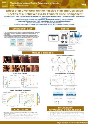
In this study, a retrieved CoCr femoral component explanted after two years of clinical service was investigated to evaluate the influence of in vivo wear on its corrosion behavior. Two representative regions were identified: a worn area located on the medial condyle and a non-worn area on the anterior surface of the prosthesis. Since the wear scars were macroscopically visible, surface characterization was performed using stereomicroscopy to document the extent and morphology of the damage.
Electrochemical characterization was conducted in phosphate-buffered saline (PBS, pH 7.4) at 37 °C to simulate physiological conditions. A custom electrochemical cell holder was fabricated by 3D printing to isolate and evaluate each selected region independently. The corrosion behavior of both zones was assessed through open circuit potential, electrochemical impedance spectroscopy, linear polarization resistance, and potentiodynamic polarization measurements.
The results provide information on the effect of in vivo wear-induced surface degradation on the passive behavior and corrosion resistance of CoCr knee implants. Differences in electrochemical response between worn and non-worn regions are discussed in terms of passive film stability and surface damage, contributing to a better understanding of tribocorrosion mechanisms in retrieved orthopedic prostheses.
Keywords
Tribocorrosion
aseptic loosening
knee prosthesis
electrochemistry
cobalt chromium alloy
Poster
IOCME-Poster-Template-6aa396de92423.pptx.pdf
Design, processing, and characterization of novel high-entropy alloys for biomedical applications by using non-toxic and low-cost alloying elements
Plastic Deformation Behavior in the Dynamic High Strain Rate of Additively Manufactured AlSi10Mg