Event submissions
Published
This submission belongs to the session S2. Acute Myeloid Leukemia of the event The 1st International Online Conference on Hematology
Published date
23 Oct, 2026
Academic Editor
 Claudio Cerchione
Citation
Roihan Awg Isa, Aida Nurul Hanim Abdul Rahman, Kimberly Fe Joibi, Noor Hayati Sabtu, Beyond Physiological Monocytes: Diagnostic Challenges in Infantile AML with KAT6A::CREBBP Fusion, in Proceedings of The 1st International Online Conference on Hematology, 28 October–30 October 2026, MDPI: Basel, Switzerland
Share
Email
Facebook
Twitter
LinkedIn
Beyond Physiological Monocytes: Diagnostic Challenges in Infantile AML with KAT6A::CREBBP Fusion

Roihan Awg Isa 1
Aida Nurul Hanim Abdul Rahman 1
Kimberly Fe Joibi 1
Noor Hayati Sabtu 1
1. Department of Pathology, Hematology, Women and Children Hospital, Likas, Sabah, Malaysia
Abstract
Introduction/Background
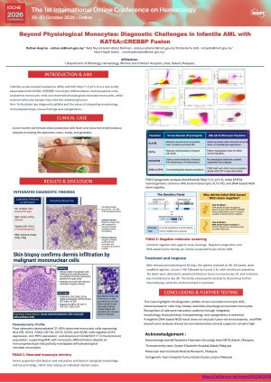
Infantile acute myeloid leukaemia (AML) with t(8;16)(p11.2;p13.3)/KAT6A::CREBBP is a rare fusion-driven AML frequently associated with monocytic differentiation and leukemia cutis. Diagnosis may be challenging because leukemic cells can resemble physiological neonatal monocytes, while routine molecular assays may not detect the underlying fusion.
Clinical Case
A one-month-old female infant presented with fever and recurrent erythematous nodules involving the abdomen, back, scalp, and genitalia. Laboratory investigations demonstrated leukocytosis, thrombocytopenia, and 25–30% circulating blasts. Morphology suggested AML with monocytic differentiation. Flow cytometry identified 31–45% abnormal monocytic cells expressing HLA-DR, CD14, CD64, CD11b, CD13, CD33, and CD36, with negative CD16, dim MPO, and a lack of CD34/CD117 in the dominant population. Partial phenotypic overlap with physiological neonatal monocytes created a diagnostic pitfall. Skin biopsy confirmed AML with monocytic differentiation. Cerebrospinal fluid asessment demonstrated CNS involvement. Conventional and FISH cytogenetic analysis demonstrated t(8;16)(p11.2;p13.3), whereas KMT2A rearrangement, common AML fusion transcripts, FLT3-ITD, and DNA-based next-generation sequencing were negative. The patient received AML-directed chemotherapy after disease persistence beyond 60 days, using an ML-DS-based dose-modified regimen, comprising Course 1 AIE followed by Course 2 AI and intrathecal Cytarabine. No blasts were detected in peripheral blood and bone marrow by day 24, and remission was maintained at the day-38 assessment. The family subsequently elected not to continue further chemotherapy while the child remained in remission.
Clinical Lessons/Conclusions
This case highlights two diagnostic pitfalls . First, leukemic monocytic populations may resemble physiological neonatal monocytes, necessitating careful assessment of population distribution and maturation architecture rather than reliance on individual markers alone. Second, the negative routine fusion screening and DNA-based NGS results do not exclude fusion-driven AML. Targeted cytogenetic studies and RNA-based fusion NGS testing should be pursued further when clinopathological findings suggest underlying fusion transcripts such as KAT6A::CREBBP.
Keywords
Keywords: Infantile AML
Flow Cytometry
KAT6A::CREBBP
Leukaemia cutis
Poster
Roihan IOCHE2026 Hematology conference E-Poster.pdf
Left Ventricular Aneurysm after MINOCA in Antiphospholipid Syndrome: A Single-Center Cohort Study
Masked Acute Promyelocytic Leukemia with Monosomy 17 and Marker Chromosome: Importance of Multimodal Molecular Diagnosis